
Search Count: 11
 |
Organism: Bacillus subtilis subsp. subtilis str. 168
Method: X-RAY DIFFRACTION Resolution:1.90 Å Release Date: 2024-09-25 Classification: LYASE Ligands: GOL, NA, CL |
 |
Semet-Derived Crystal Structure Of The N-Terminal Beta-Hairpin Docking (Bhd) Domain Of The Aerj Halogenase, From The Aeruginosin Biosynthetic Assembly Line
Organism: Microcystis aeruginosa nies-98
Method: X-RAY DIFFRACTION Resolution:1.68 Å Release Date: 2024-03-06 Classification: PROTEIN BINDING Ligands: CL |
 |
Native Crystal Structure Of The N-Terminal Beta-Hairpin Docking (Bhd) Domain Of The Aerj Halogenase, From The Aeruginosin Biosynthetic Assembly Line
Organism: Microcystis aeruginosa nies-98
Method: X-RAY DIFFRACTION Resolution:1.46 Å Release Date: 2024-03-06 Classification: PROTEIN BINDING Ligands: CL |
 |
Crystal Structure Of Pksd, The Trans-Acting Acyl Hydrolase Domain From The Bacillaene Trans-At Pks (Semet Derivative)
Organism: Bacillus subtilis subsp. subtilis str. 168
Method: X-RAY DIFFRACTION Resolution:1.96 Å Release Date: 2023-08-09 Classification: HYDROLASE Ligands: ZN |
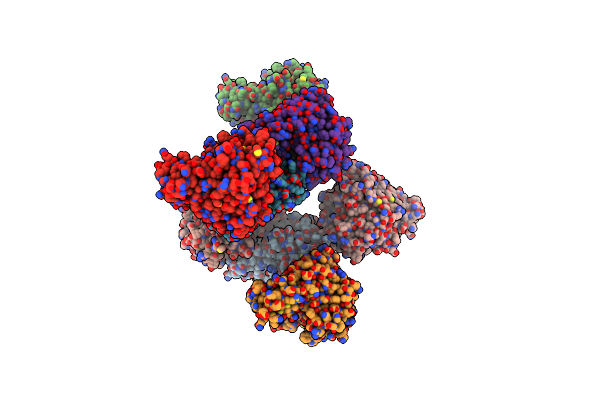 |
Crystal Structure Of Pksd, The Trans-Acting Acyl Hydrolase Domain From The Bacillaene Trans-At Pks (Native)
Organism: Bacillus subtilis subsp. subtilis str. 168
Method: X-RAY DIFFRACTION Resolution:2.20 Å Release Date: 2023-08-09 Classification: HYDROLASE Ligands: ZN |
 |
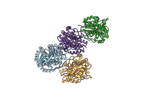
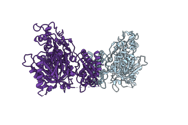
Crystal Structure Of The Epimerization Domain From The Third Module Of Tyrocidine Synthetase B, Tycb3(E)
Organism: Brevibacillus parabrevis
Method: X-RAY DIFFRACTION Resolution:2.40 Å Release Date: 2020-11-18 Classification: BIOSYNTHETIC PROTEIN |
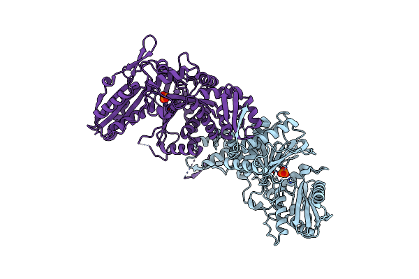 |
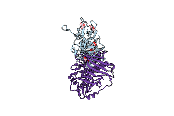
Molecular Basis For Condensation Domain-Mediated Chain Release From The Enacyloxin Polyketide Synthase
Organism: Burkholderia ambifaria (strain atcc baa-244 / ammd)
Method: X-RAY DIFFRACTION Resolution:2.00 Å Release Date: 2019-02-27 Classification: BIOSYNTHETIC PROTEIN Ligands: PO4 |
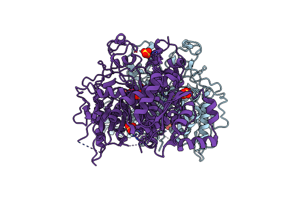 |
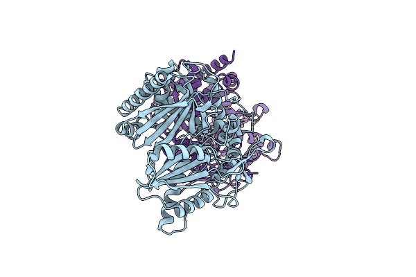
Crystal Structure Of The Second Ketosynthase From The Bacillaene Polyketide Synthase
Organism: Bacillus subtilis subsp. subtilis
Method: X-RAY DIFFRACTION Resolution:1.95 Å Release Date: 2014-02-19 Classification: TRANSFERASE Ligands: SO4 |
 |
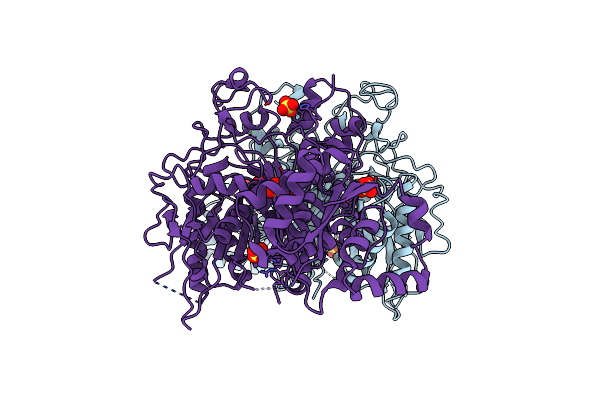
Crystal Structure Of The Second Ketosynthase From The Bacillaene Polyketide Synthase Bound To Its Natural Intermediate
Organism: Bacillus subtilis subsp. subtilis
Method: X-RAY DIFFRACTION Resolution:2.30 Å Release Date: 2014-02-19 Classification: TRANSFERASE |
 |
Crystal Structure Of The Second Ketosynthase From The Bacillaene Polyketide Synthase Bound To A Hexanoyl Substrate Mimic
Organism: Bacillus subtilis subsp. subtilis
Method: X-RAY DIFFRACTION Resolution:2.89 Å Release Date: 2014-02-19 Classification: TRANSFERASE Ligands: SO4 |
 |
Crystal Structure Of Quinone Reductase 2 In Complex With The Indolequinone Mac627
Organism: Homo sapiens
Method: X-RAY DIFFRACTION Resolution:2.00 Å Release Date: 2011-05-11 Classification: OXIDOREDUCTASE/OXIDOREDUCTASE INHIBITOR Ligands: ZN, FAD, O73 |