
Search Count: 6
 |
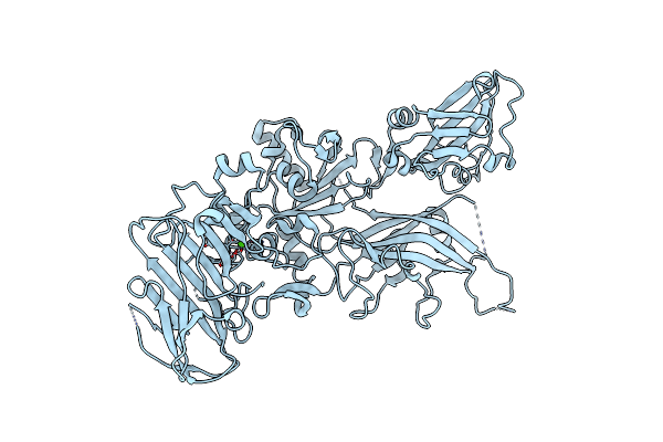
Organism: Bacillus anthracis
Method: X-RAY DIFFRACTION Resolution:2.00 Å Release Date: 2012-02-15 Classification: TOXIN Ligands: CA |
 |
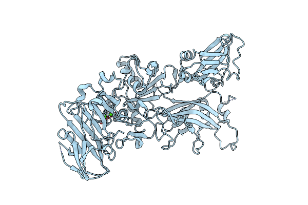
Organism: Bacillus anthracis
Method: X-RAY DIFFRACTION Resolution:2.85 Å Release Date: 2012-02-15 Classification: TOXIN Ligands: CA |
 |
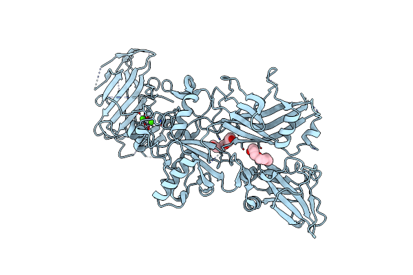
Organism: Bacillus anthracis
Method: X-RAY DIFFRACTION Resolution:2.10 Å Release Date: 2012-02-15 Classification: TOXIN Ligands: CA |
 |
Organism: Bacillus anthracis
Method: X-RAY DIFFRACTION Resolution:2.10 Å Release Date: 2012-02-15 Classification: PROTEIN BINDING Ligands: CA, PG4 |
 |
Organism: Bacillus anthracis
Method: X-RAY DIFFRACTION Resolution:3.13 Å Release Date: 2012-01-18 Classification: TOXIN Ligands: CA |
 |
Organism: Bacillus anthracis
Method: X-RAY DIFFRACTION Resolution:1.70 Å Release Date: 2010-08-11 Classification: TOXIN Ligands: CA, PG4 |