Planned Maintenance: Some services may turn out to be unavailable from 15th January, 2026 to 16th January, 2026. We apologize for the inconvenience!

Planned Maintenance: Some services may turn out to be unavailable from 15th January, 2026 to 16th January, 2026. We apologize for the inconvenience!
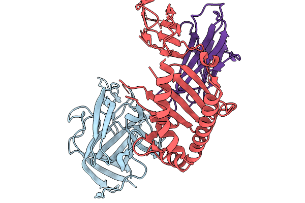 |
Organism: Homo sapiens, Human astrovirus 6
Method: X-RAY DIFFRACTION Release Date: 2025-11-19 Classification: IMMUNE SYSTEM |
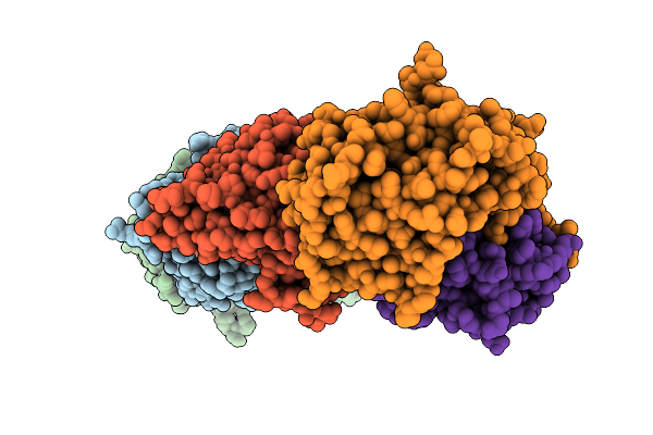 |
Organism: Human astrovirus 2, Homo sapiens
Method: X-RAY DIFFRACTION Release Date: 2025-11-19 Classification: IMMUNE SYSTEM |
 |
Crystal Structure Of Fab Ms-1805 In Complex With Npna3 Peptide From Circumsporozoite Protein
Organism: Homo sapiens, Plasmodium falciparum 3d7
Method: X-RAY DIFFRACTION Release Date: 2025-10-01 Classification: IMMUNE SYSTEM |
 |
Crystal Structure Of Fab 7088 In Complex With Npna3 Peptide From Circumsporozoite Protein
Organism: Homo sapiens, Plasmodium falciparum 3d7
Method: X-RAY DIFFRACTION Release Date: 2025-10-01 Classification: IMMUNE SYSTEM |
 |
Crystal Structure Of Fab Ms-1805 In Complex With N-Terminal Junction Peptide From Circumsporozoite Protein
Organism: Homo sapiens, Plasmodium falciparum 3d7
Method: X-RAY DIFFRACTION Release Date: 2025-10-01 Classification: IMMUNE SYSTEM Ligands: CIT |
 |
Crystal Structure Of Fab 7088 In Complex With N-Terminal Junction Peptide From Circumsporozoite Protein
Organism: Homo sapiens, Plasmodium falciparum 3d7
Method: X-RAY DIFFRACTION Release Date: 2025-10-01 Classification: IMMUNE SYSTEM |
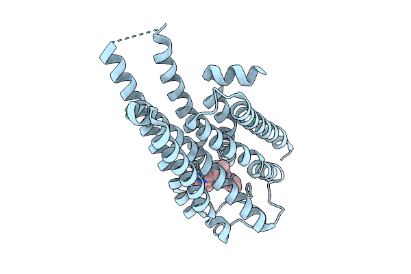 |
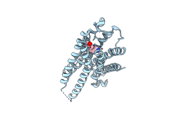
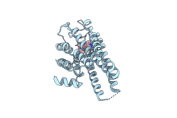
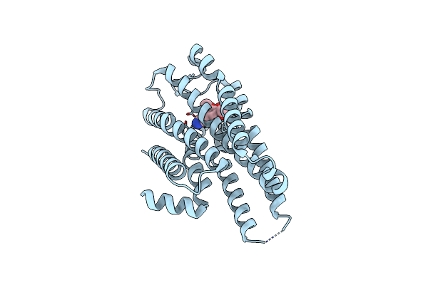
Local Refinement Of Drd2 Bound To Lsd In Complex With A Mini-Goa And Scfv16 Obtained By Cryo-Electron Microscopy (Cryoem)
Organism: Escherichia coli, Homo sapiens
Method: ELECTRON MICROSCOPY Release Date: 2025-09-17 Classification: MEMBRANE PROTEIN Ligands: 7LD |
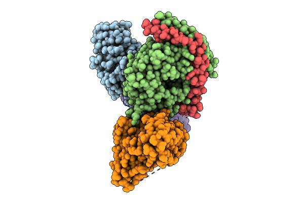 |
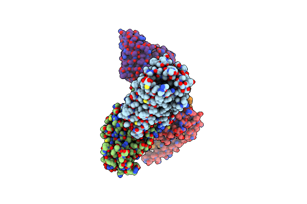
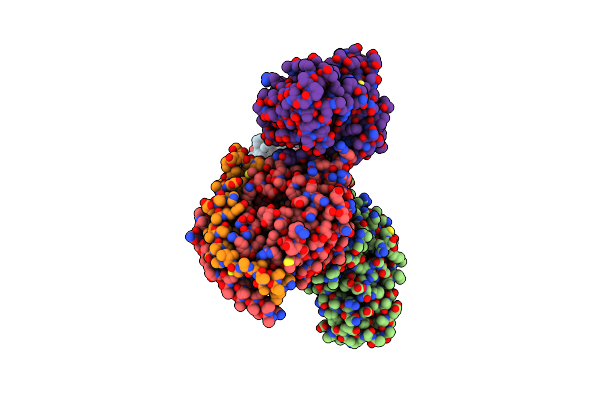
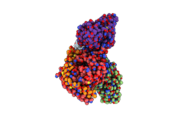
Global Reconstruction Of Drd2 Bound To Lsd In Complex With A Mini-Goa And Scfv16 Obtained By Cryo-Electron Microscopy (Cryoem)
Organism: Homo sapiens, Escherichia coli, Mus musculus
Method: ELECTRON MICROSCOPY Release Date: 2025-09-17 Classification: MEMBRANE PROTEIN Ligands: 7LD |
 |
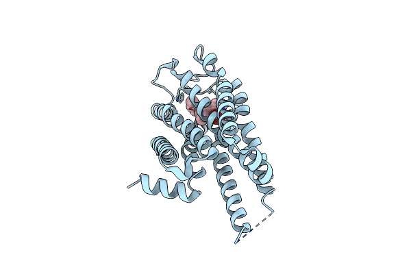
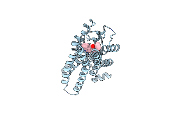
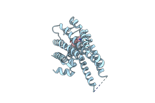
Local Refinement Of 5-Ht2Ar Bound To 5-Ht In Complex With A Mini-Gq Protein And Scfv16 Obtained By Cryo-Electron Microscopy (Cryoem)
Organism: Homo sapiens
Method: ELECTRON MICROSCOPY Release Date: 2025-04-02 Classification: MEMBRANE PROTEIN Ligands: SRO |
 |
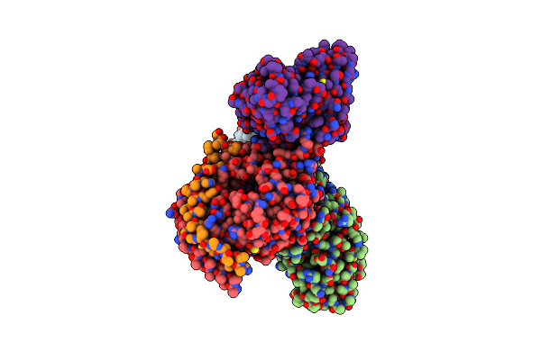
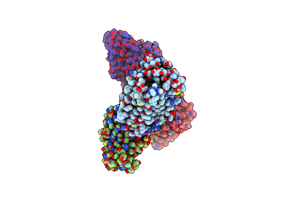
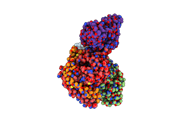
Global Reconstruction 5-Ht2Ar Bound To 5-Ht In Complex With A Mini-Gq Protein And Scfv16 Obtained By Cryo-Electron Microscopy (Cryoem)
Organism: Homo sapiens
Method: ELECTRON MICROSCOPY Resolution:3.27 Å Release Date: 2025-04-02 Classification: MEMBRANE PROTEIN Ligands: SRO |
 |
Local Refinement Of 5-Ht2Ar Bound To 2-Bromo-Lsd In Complex With A Mini-Gq Protein And Scfv16 Obtained By Cryo-Electron Microscopy (Cryoem)
Organism: Homo sapiens
Method: ELECTRON MICROSCOPY Resolution:3.37 Å Release Date: 2025-04-02 Classification: MEMBRANE PROTEIN Ligands: A1AFU |
 |
Global Reconstruction Of 5-Ht2Ar Bound To 2-Bromo-Lsd In Complex With A Mini-Gq Protein And Scfv16 Obtained By Cryo-Electron Microscopy (Cryoem)
Organism: Homo sapiens
Method: ELECTRON MICROSCOPY Resolution:3.38 Å Release Date: 2025-04-02 Classification: MEMBRANE PROTEIN Ligands: A1AFU |
 |
Local Refinement Of 5-Ht2Ar Bound To Dmt In Complex With A Mini-Gq Protein And Scfv16 Obtained By Cryo-Electron Microscopy (Cryoem)
Organism: Homo sapiens
Method: ELECTRON MICROSCOPY Resolution:3.38 Å Release Date: 2025-04-02 Classification: MEMBRANE PROTEIN Ligands: A1AFV |
 |
Global Reconstruction Of 5-Ht2Ar Bound To Dmt In Complex With A Mini-Gq Protein And Scfv16 Obtained By Cryo-Electron Microscopy (Cryoem)
Organism: Homo sapiens
Method: ELECTRON MICROSCOPY Resolution:3.21 Å Release Date: 2025-04-02 Classification: MEMBRANE PROTEIN Ligands: A1AFV |
 |
Local Refinement Of 5-Ht2Ar Bound To Lsd In Complex With A Mini-Gq Protein And Scfv16 Obtained By Cryo-Electron Microscopy (Cryoem)
Organism: Homo sapiens
Method: ELECTRON MICROSCOPY Release Date: 2025-04-02 Classification: MEMBRANE PROTEIN Ligands: 7LD |
 |
Global Reconstruction Of 5-Ht2Ar Bound To Lsd In Complex With A Mini-Gq Protein And Scfv16 Obtained By Cryo-Electron Microscopy (Cryoem)
Organism: Homo sapiens
Method: ELECTRON MICROSCOPY Release Date: 2025-04-02 Classification: MEMBRANE PROTEIN Ligands: 7LD |
 |
Local Refinement Of 5-Ht2Ar Bound To Mescaline In Complex With A Mini-Gq Protein And Scfv16 Obtained By Cryo-Electron Microscopy (Cryoem)
Organism: Homo sapiens
Method: ELECTRON MICROSCOPY Release Date: 2025-04-02 Classification: MEMBRANE PROTEIN Ligands: A1AFW |
 |
Global Reconstruction Of 5-Ht2Ar Bound To Mescaline In Complex With A Mini-Gq Protein And Scfv16 Obtained By Cryo-Electron Microscopy (Cryoem)
Organism: Homo sapiens
Method: ELECTRON MICROSCOPY Release Date: 2025-04-02 Classification: MEMBRANE PROTEIN Ligands: A1AFW |
 |
Local Refinement Of 5-Ht2Ar Bound To Psilocin In Complex With A Mini-Gq And Scfv16 Obtained By Cryo-Electron Microscopy (Cryoem)
Organism: Homo sapiens
Method: ELECTRON MICROSCOPY Resolution:2.72 Å Release Date: 2025-04-02 Classification: MEMBRANE PROTEIN Ligands: 91Q |
 |
Global Reconstruction Of 5-Ht2Ar Bound To Psilocin In Complex With A Mini-Gq Protein And Scfv16 Obtained By Cryo-Electron Microscopy (Cryoem)
Organism: Homo sapiens
Method: ELECTRON MICROSCOPY Resolution:2.54 Å Release Date: 2025-04-02 Classification: MEMBRANE PROTEIN Ligands: 91Q |