
Search Count: 171
 |
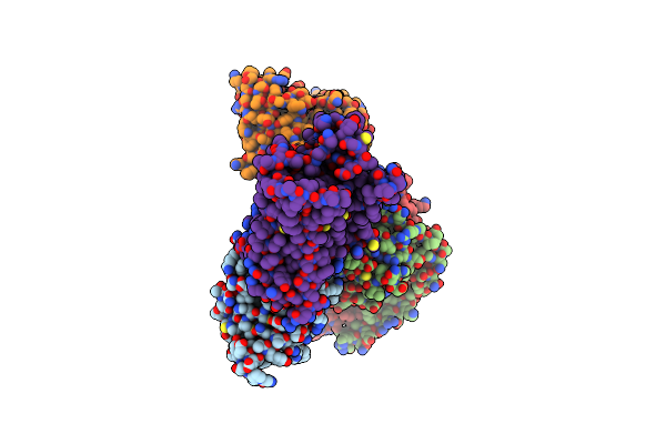
Organism: Synthetic construct, Ovis aries
Method: ELECTRON MICROSCOPY Release Date: 2023-03-22 Classification: MEMBRANE PROTEIN |
 |
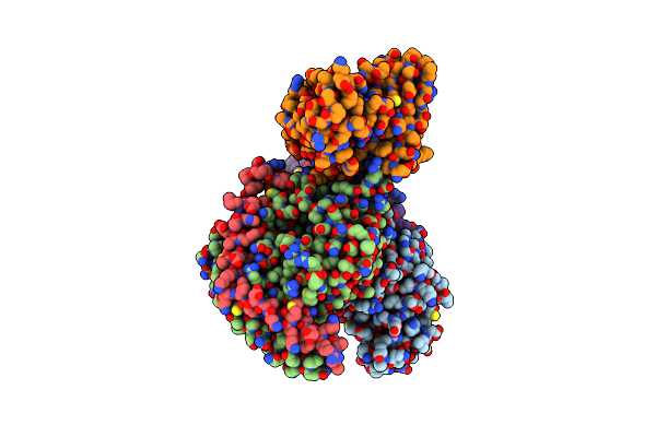
Cryo-Em Structure Of The Alpha2A Adrenergic Receptor Goa Signaling Complex Bound To A G Protein Biased Agonist
Organism: Homo sapiens, Mus musculus
Method: ELECTRON MICROSCOPY Release Date: 2022-09-28 Classification: MEMBRANE PROTEIN Ligands: W96 |
 |
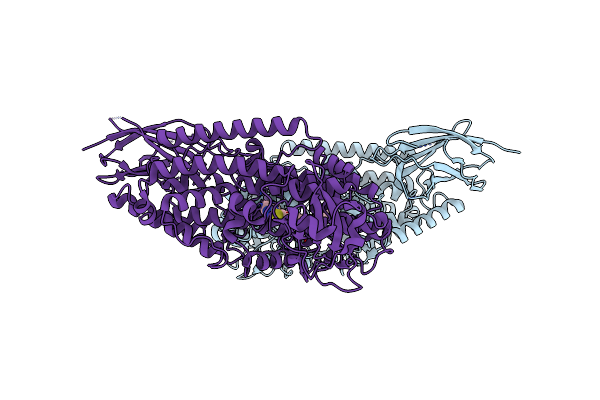
Cryo-Em Structure Of The Alpha2A Adrenergic Receptor Goa Signaling Complex Bound To A Biased Agonist
Organism: Homo sapiens, Mus musculus
Method: ELECTRON MICROSCOPY Release Date: 2022-09-28 Classification: MEMBRANE PROTEIN Ligands: W58 |
 |
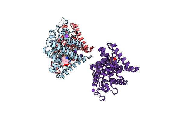
Organism: Homo sapiens
Method: X-RAY DIFFRACTION Resolution:2.44 Å Release Date: 2022-01-19 Classification: OXIDOREDUCTASE/OXIDOREDUCTASE INHIBITOR Ligands: MN, XRP |
 |
Organism: Homo sapiens
Method: ELECTRON MICROSCOPY Release Date: 2021-04-21 Classification: LIPID BINDING PROTEIN |
 |
Crystal Structure Of Streptogramin A Acetyltransferase Vata From Staphylococcus Aureus In Complex With Streptogramin Analog F1037 (47)
Organism: Staphylococcus aureus
Method: X-RAY DIFFRACTION Resolution:3.05 Å Release Date: 2020-11-18 Classification: TRANSFERASE Ligands: SXA, O7S, PO4, SO4, CL, MG |
 |
Crystal Structure Of Streptogramin A Acetyltransferase Vata From Staphylococcus Aureus In Complex With Streptogramin Analog F0224 (46)
Organism: Staphylococcus aureus
Method: X-RAY DIFFRACTION Resolution:2.70 Å Release Date: 2020-11-18 Classification: TRANSFERASE Ligands: O7V, PO4, SXA, CL, MG |
 |
Organism: Escherichia coli, Streptomyces virginiae
Method: ELECTRON MICROSCOPY Release Date: 2020-06-17 Classification: RIBOSOME Ligands: O7V |
 |
Organism: Escherichia coli
Method: ELECTRON MICROSCOPY Release Date: 2020-06-17 Classification: RIBOSOME Ligands: O7S |
 |
Organism: Escherichia coli
Method: ELECTRON MICROSCOPY Release Date: 2020-06-17 Classification: RIBOSOME Ligands: O7V |
 |
Organism: Escherichia coli
Method: ELECTRON MICROSCOPY Release Date: 2020-06-17 Classification: RIBOSOME Ligands: O7Y |
 |
Organism: Escherichia coli
Method: ELECTRON MICROSCOPY Release Date: 2020-06-17 Classification: RIBOSOME Ligands: O8D |
 |
Organism: Escherichia coli
Method: ELECTRON MICROSCOPY Release Date: 2020-06-17 Classification: RIBOSOME Ligands: O8J |
 |
Organism: Escherichia coli
Method: ELECTRON MICROSCOPY Release Date: 2020-06-17 Classification: RIBOSOME Ligands: O8P |
 |
Organism: Escherichia coli
Method: ELECTRON MICROSCOPY Release Date: 2020-06-17 Classification: RIBOSOME Ligands: O8S |
 |
Organism: Escherichia coli
Method: ELECTRON MICROSCOPY Release Date: 2020-06-17 Classification: RIBOSOME Ligands: O8V |
 |
Organism: Escherichia coli, Streptomyces virginiae
Method: ELECTRON MICROSCOPY Release Date: 2020-06-17 Classification: RIBOSOME Ligands: O7S |
 |
Organism: Homo sapiens
Method: X-RAY DIFFRACTION Resolution:3.20 Å Release Date: 2019-03-20 Classification: TRANSCRIPTION Ligands: K, G7J, BR |
 |
Crystal Structure Of Carbonmonoxy Hemoglobin S (Liganded Sickle Cell Hemoglobin) Complexed With Gbt Compound 31
Organism: Homo sapiens
Method: X-RAY DIFFRACTION Resolution:1.95 Å Release Date: 2017-02-22 Classification: OXYGEN TRANSPORT Ligands: HEM, CMO, 7SJ |
 |
Crystal Structure Of Carbonmonoxy Hemoglobin S (Liganded Sickle Cell Hemoglobin) Complexed With Gbt Compound 6
Organism: Homo sapiens
Method: X-RAY DIFFRACTION Resolution:2.05 Å Release Date: 2017-02-22 Classification: OXYGEN TRANSPORT Ligands: HEM, CMO, 86M |