
Search Count: 90
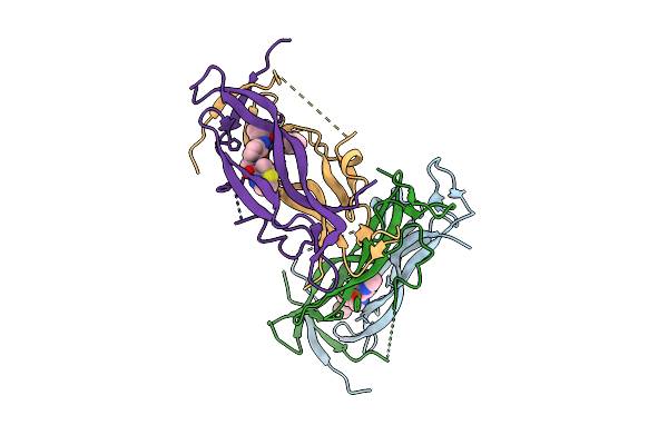 |
Organism: Homo sapiens
Method: X-RAY DIFFRACTION Resolution:3.15 Å Release Date: 2025-10-15 Classification: CYTOKINE Ligands: A1CXF |
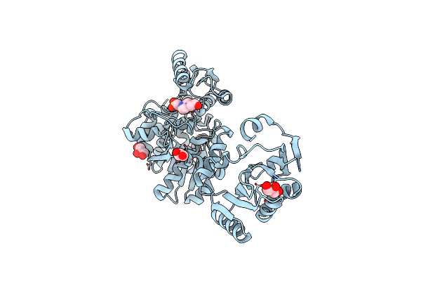 |
Organism: Escherichia coli
Method: X-RAY DIFFRACTION Resolution:1.87 Å Release Date: 2025-04-09 Classification: LYASE Ligands: GOL, PGE, EPE |
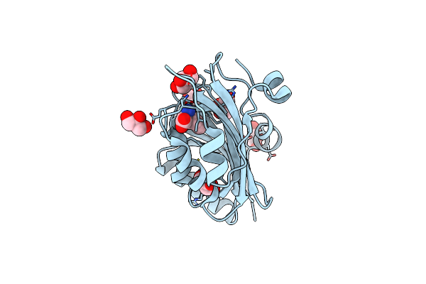 |
Crystal Structure Of E. Coli Threonine Dehydratase Regulatory Domain In Complex With Isoleucine
Organism: Escherichia coli
Method: X-RAY DIFFRACTION Resolution:1.60 Å Release Date: 2025-04-09 Classification: LYASE Ligands: ILE, GOL, ACT |
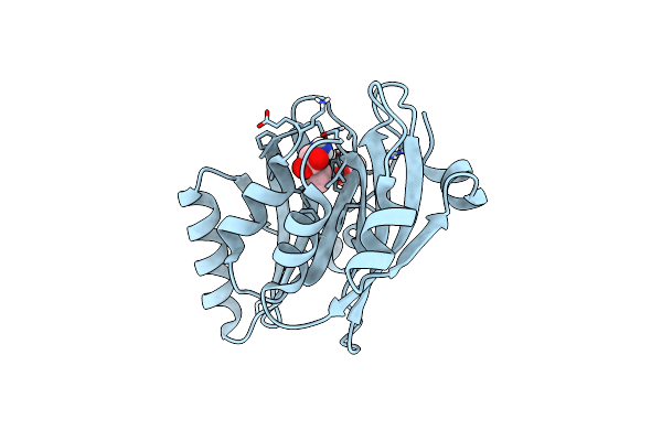 |
Crystal Structure Of E. Coli Threonine Dehydratase Regulatory Domain F352A Mutant In Complex With Isoleucine
Organism: Escherichia coli
Method: X-RAY DIFFRACTION Resolution:1.22 Å Release Date: 2025-04-09 Classification: LYASE Ligands: ILE, GOL |
 |
Organism: Staphylococcus aureus
Method: X-RAY DIFFRACTION Resolution:2.80 Å Release Date: 2025-04-09 Classification: LYASE |
 |
Four Subunit Cytochrome B-C1 Complex From Rhodobacter Sphaeroides In Native Nanodiscs - Consensus Refinement In The B-B Conformation
Organism: Cereibacter sphaeroides 2.4.1
Method: ELECTRON MICROSCOPY Release Date: 2023-03-15 Classification: OXIDOREDUCTASE Ligands: FES, PEE, HEM, HEC, U10 |
 |
Four Subunit Cytochrome B-C1 Complex From Rhodobacter Sphaeroides In Native Nanodiscs - Focussed Refinement In The B-C Conformation
Organism: Cereibacter sphaeroides 2.4.1
Method: ELECTRON MICROSCOPY Release Date: 2023-03-15 Classification: OXIDOREDUCTASE Ligands: FES, PEE, HEM, HEC, U10 |
 |
Organism: Homo sapiens
Method: X-RAY DIFFRACTION Resolution:1.99 Å Release Date: 2022-11-30 Classification: CELL CYCLE Ligands: 799 |
 |
Organism: Homo sapiens
Method: X-RAY DIFFRACTION Resolution:2.46 Å Release Date: 2022-11-30 Classification: CELL CYCLE Ligands: X3W |
 |
Organism: Homo sapiens
Method: X-RAY DIFFRACTION Resolution:2.68 Å Release Date: 2022-11-30 Classification: CELL CYCLE Ligands: X3R |
 |
Organism: Homo sapiens
Method: X-RAY DIFFRACTION Resolution:2.48 Å Release Date: 2022-11-30 Classification: CELL CYCLE Ligands: X3N |
 |
Organism: Rhodopseudomonas palustris atcc 17001
Method: ELECTRON MICROSCOPY Release Date: 2022-10-12 Classification: PHOTOSYNTHESIS Ligands: BCL, IRM |
 |
Organism: Rhodopseudomonas palustris
Method: ELECTRON MICROSCOPY Release Date: 2022-10-05 Classification: PHOTOSYNTHESIS Ligands: BCL, IRM |
 |
Organism: Rhodopseudomonas palustris
Method: ELECTRON MICROSCOPY Release Date: 2022-10-05 Classification: PHOTOSYNTHESIS Ligands: BCL, ZE0 |
 |
Organism: Rhodopseudomonas palustris
Method: ELECTRON MICROSCOPY Release Date: 2022-10-05 Classification: PHOTOSYNTHESIS Ligands: BCL, IRM |
 |
Cryo-Em Structure (Model_1A) Of The Rc-Dlh Complex From Gemmatimonas Phototrophica At 2.4 A
Organism: Gemmatimonas phototrophica
Method: ELECTRON MICROSCOPY Release Date: 2022-03-02 Classification: MEMBRANE PROTEIN Ligands: BCL, LMT, V7N, 0V9, HEC, NDG, V75, CD4, PGW, MQ8, BPH, FE, CRT, V7B, UYH |
 |
Cryo-Em Structure (Model_2A) Of The Rc-Dlh Complex From Gemmatimonas Phototrophica At 2.5 A
Organism: Gemmatimonas phototrophica
Method: ELECTRON MICROSCOPY Release Date: 2022-03-02 Classification: MEMBRANE PROTEIN Ligands: BCL, LMT, V7N, HEC, V75, NDG, 0V9, CD4, PGW, MQ8, V7B, BPH, FE, CRT, UYH |
 |
Cryo-Em Structure Of The Rc-Dlh Complex (Model_1B) From Gemmatimonas Phototrophica At 2.47 A
Organism: Gemmatimonas phototrophica
Method: ELECTRON MICROSCOPY Release Date: 2022-03-02 Classification: MEMBRANE PROTEIN Ligands: BCL, LMT, V7N, 0V9, HEC, V75, NDG, PGW, CD4, BPH, MQ8, FE, CRT, V7B, UYH |
 |
Cryo-Em Structure (Model_2B) Of The Rc-Dlh Complex From Gemmatimonas Phototrophica At 2.44 A
Organism: Gemmatimonas phototrophica
Method: ELECTRON MICROSCOPY Release Date: 2022-03-02 Classification: MEMBRANE PROTEIN Ligands: BCL, LMT, V7N, HEC, NDG, V75, PGW, 0V9, CD4, BPH, MQ8, FE, CRT, V7B, UYH |
 |
Cryo-Em Structure Of The Dimeric Rhodobacter Sphaeroides Rc-Lh1 Core Complex At 2.9 A: The Structural Basis For Dimerisation
Organism: Cereibacter sphaeroides 2.4.1
Method: ELECTRON MICROSCOPY Release Date: 2021-11-24 Classification: PHOTOSYNTHESIS Ligands: BCL, SP2, 3PE, CD4, UQ1, U10, BPH, SQD, LMT, FE |