
Search Count: 14
 |
Organism: Homo sapiens
Method: ELECTRON MICROSCOPY Release Date: 2023-11-08 Classification: PROTEIN FIBRIL |
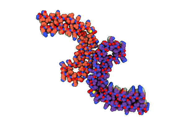 |
Structure Of Tau Filaments Type Ii From Subacute Sclerosing Panencephalitis
Organism: Homo sapiens
Method: ELECTRON MICROSCOPY Release Date: 2023-11-08 Classification: PROTEIN FIBRIL |
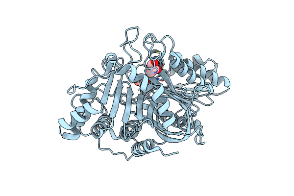 |
Organism: Streptomyces antibioticus
Method: X-RAY DIFFRACTION Resolution:2.49 Å Release Date: 2021-04-28 Classification: HYDROLASE Ligands: MES, GOL |
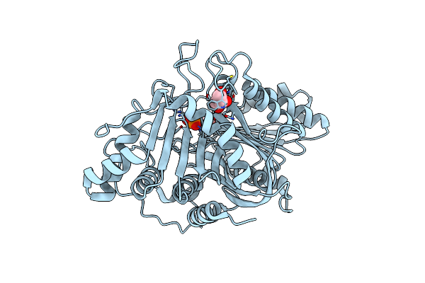 |
Organism: Streptomyces antibioticus
Method: X-RAY DIFFRACTION Resolution:2.01 Å Release Date: 2021-04-28 Classification: HYDROLASE Ligands: MES, PO4, GOL |
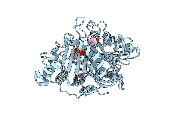 |
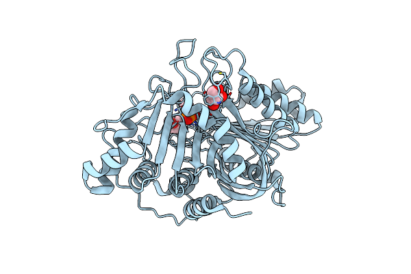
Organism: Streptomyces antibioticus
Method: X-RAY DIFFRACTION Resolution:2.21 Å Release Date: 2021-04-28 Classification: HYDROLASE Ligands: MES, GOL, VHY |
 |
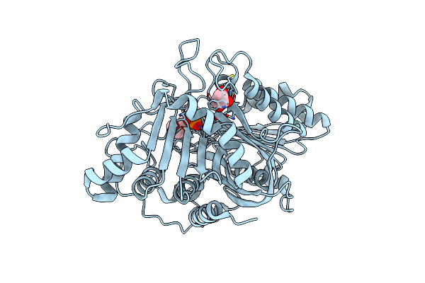
Phospholipase D Engineered Mutant Bound To Phosphatidic Acid (30 Minute Soak)
Organism: Streptomyces antibioticus
Method: X-RAY DIFFRACTION Resolution:2.42 Å Release Date: 2021-04-28 Classification: HYDROLASE Ligands: MES, GOL, VHY |
 |
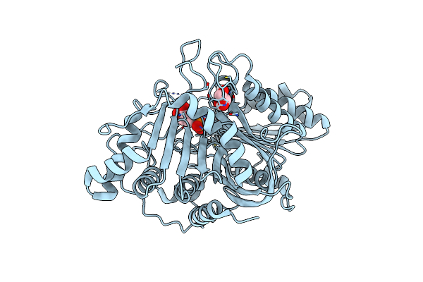
Organism: Streptomyces antibioticus
Method: X-RAY DIFFRACTION Resolution:1.99 Å Release Date: 2021-04-28 Classification: HYDROLASE Ligands: MES, GOL, VHY |
 |
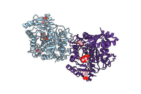
Phospholipase D Engineered Mutant (Tnyr) Inactive Enzyme (H168A) Bound To 1-Inositol Phosphate
Organism: Streptomyces antibioticus
Method: X-RAY DIFFRACTION Resolution:2.50 Å Release Date: 2021-04-28 Classification: LIPID BINDING PROTEIN Ligands: IPD, MES, GOL |
 |
Phospholipase D Engineered Mutant (Tnyr) H442 Covalent Adduct With 1-Inositol Phosphate
Organism: Streptomyces antibioticus
Method: X-RAY DIFFRACTION Resolution:2.30 Å Release Date: 2021-04-28 Classification: HYDROLASE Ligands: GOL, MES, IPD |
 |
Organism: Aeropyrum pernix
Method: X-RAY DIFFRACTION Resolution:2.07 Å Release Date: 2012-05-16 Classification: TRANSFERASE Ligands: PLP, MPD |
 |
Crystal Structure Of The K127A Mutant Of O-Phosphoserine Sulfhydrylase Complexed With External Schiff Base Of Pyridoxal 5'-Phosphate With O-Phospho-L-Serine
Organism: Aeropyrum pernix
Method: X-RAY DIFFRACTION Resolution:2.07 Å Release Date: 2012-05-16 Classification: TRANSFERASE Ligands: PLP, SEP, MPD |
 |
Crystal Structure Of The K127A Mutant Of O-Phosphoserine Sulfhydrylase Complexed With External Schiff Base Of Pyridoxal 5'-Phosphate With O-Acetyl-L-Serine
Organism: Aeropyrum pernix
Method: X-RAY DIFFRACTION Resolution:2.09 Å Release Date: 2012-05-16 Classification: TRANSFERASE Ligands: PLP, OAS, MPD |
 |
Organism: Streptomyces antibioticus
Method: X-RAY DIFFRACTION Resolution:2.50 Å Release Date: 2007-12-25 Classification: HYDROLASE Ligands: MES |
 |
Crystal Structure Of H168A Mutant Of Phospholipase D From Streptomyces Antibioticus, As A Complex With Phosphatidylcholine
Organism: Streptomyces antibioticus
Method: X-RAY DIFFRACTION Resolution:2.30 Å Release Date: 2007-12-25 Classification: HYDROLASE Ligands: MES, PD7 |