
Search Count: 37
 |
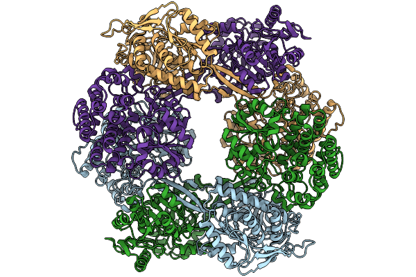
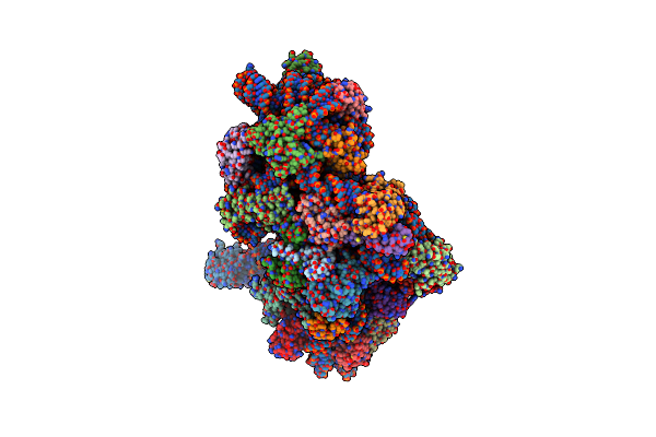
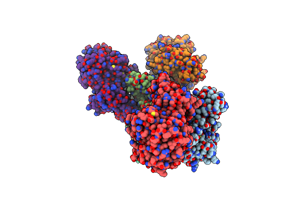
Organism: Escherichia coli k-12
Method: ELECTRON MICROSCOPY Release Date: 2025-10-01 Classification: BIOSYNTHETIC PROTEIN |
 |
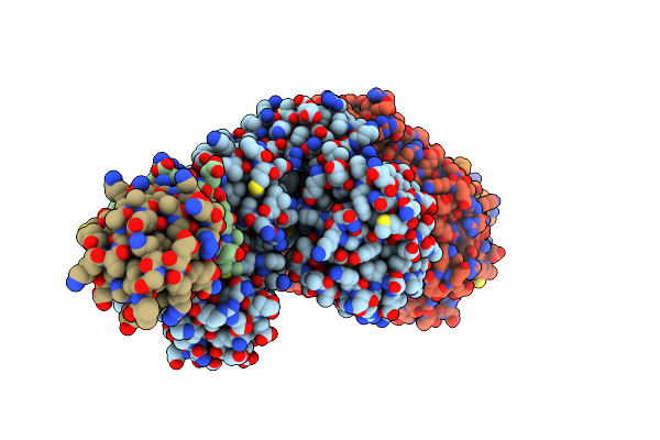
Organism: Halomonas eurihalina
Method: ELECTRON MICROSCOPY Release Date: 2025-10-01 Classification: BIOSYNTHETIC PROTEIN |
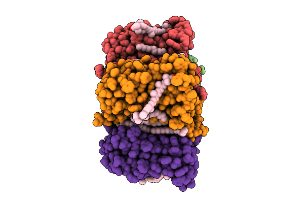 |
Cryo-Em Structure Of The Zeaxanthin-Bound Light-Driven Proton Pumping Rhodopsin, Nm-R1
Organism: Nonlabens marinus s1-08
Method: ELECTRON MICROSCOPY Release Date: 2025-07-30 Classification: MEMBRANE PROTEIN Ligands: RET, R16, D12, K3I |
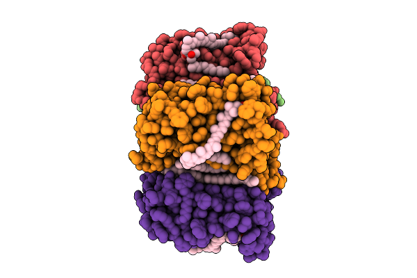 |
Cryo-Em Structure Of The Myxol-Bound Light-Driven Proton Pumping Rhodopsin, Nm-R1
Organism: Nonlabens marinus s1-08
Method: ELECTRON MICROSCOPY Release Date: 2025-07-30 Classification: MEMBRANE PROTEIN Ligands: RET, R16, D12, A1L4O |
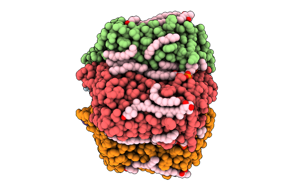 |
Cryo-Em Structure Of The Myxol-Bound Light-Driven Chloride Ion-Pumping Rhodopsin, Nm-R3
Organism: Nonlabens marinus s1-08
Method: ELECTRON MICROSCOPY Release Date: 2025-07-30 Classification: MEMBRANE PROTEIN Ligands: RET, A1L4O, CL, PC1, PLC, R16, 8K6, D12, C14, D10 |
 |
Cryo-Em Structure Of The Light-Driven Chloride Ion-Pumping Rhodopsin, Nm-R3
Organism: Nonlabens marinus s1-08
Method: ELECTRON MICROSCOPY Release Date: 2025-07-30 Classification: MEMBRANE PROTEIN Ligands: RET, CL, PC1, PLC, D12, R16, 8K6, C14 |
 |
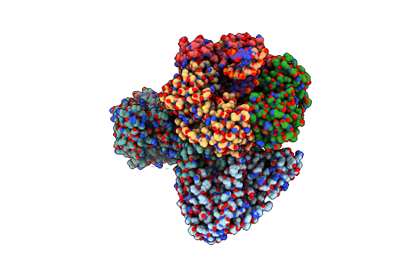
Organism: Pyrococcus furiosus
Method: ELECTRON MICROSCOPY Release Date: 2025-01-15 Classification: TRANSLATION |
 |
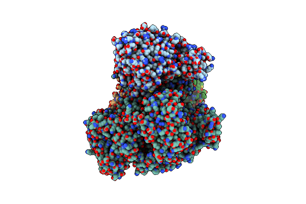
Organism: Pyrococcus furiosus
Method: ELECTRON MICROSCOPY Release Date: 2025-01-15 Classification: TRANSLATION |
 |
Organism: Pyrococcus furiosus
Method: ELECTRON MICROSCOPY Release Date: 2025-01-15 Classification: TRANSLATION |
 |
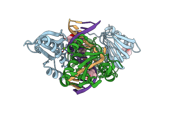
Organism: Thermococcus kodakarensis (strain atcc baa-918 / jcm 12380 / kod1)
Method: X-RAY DIFFRACTION Resolution:2.45 Å Release Date: 2021-10-06 Classification: DNA BINDING PROTEIN/REPLICATION |
 |
Organism: Thermococcus kodakarensis (strain atcc baa-918 / jcm 12380 / kod1), Thermococcus kodakarensis, Synthetic construct
Method: ELECTRON MICROSCOPY Release Date: 2020-08-05 Classification: REPLICATION/DNA Ligands: FE, ZN |
 |
Organism: Thermococcus kodakarensis kod1, Thermococcus kodakarensis, Synthetic construct
Method: ELECTRON MICROSCOPY Release Date: 2020-08-05 Classification: REPLICATION/DNA Ligands: FE, ZN |
 |
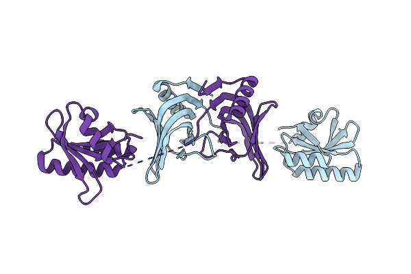
Crystal Structure Of Archaeal Ribosomal Protein Ap1, Apelota, And Gtp-Bound Aef1A Complex
Organism: Aeropyrum pernix k1
Method: X-RAY DIFFRACTION Resolution:3.00 Å Release Date: 2019-11-13 Classification: TRANSLATION Ligands: GTP, MG, NA |
 |
Crystal Structure Of The Novel Lesion-Specific Endonuclease Pfuendoq From Pyrococcus Furiosus
Organism: Pyrococcus furiosus
Method: X-RAY DIFFRACTION Resolution:2.50 Å Release Date: 2018-04-04 Classification: DNA BINDING PROTEIN Ligands: ZN, SM |
 |
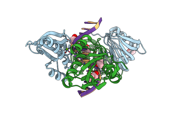
Organism: Thermococcus kodakarensis kod1, Synthetic construct
Method: X-RAY DIFFRACTION Resolution:2.40 Å Release Date: 2016-11-02 Classification: HYDROLASE/DNA Ligands: MG, MPD |
 |
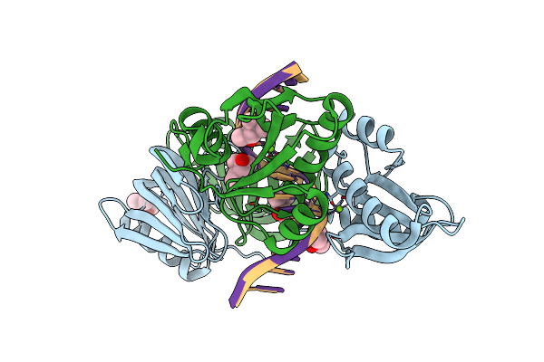
Organism: Thermococcus kodakarensis kod1, Synthetic construct
Method: X-RAY DIFFRACTION Resolution:2.80 Å Release Date: 2016-11-02 Classification: HYDROLASE/DNA Ligands: MPD, MG |
 |
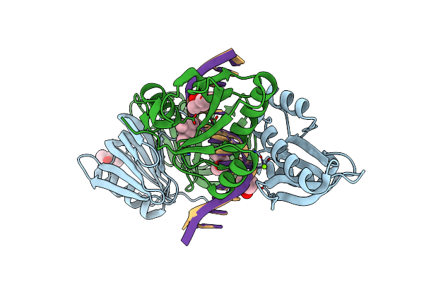
Organism: Thermococcus kodakarensis kod1, Synthetic construct
Method: X-RAY DIFFRACTION Resolution:2.60 Å Release Date: 2016-11-02 Classification: HYDROLASE/DNA Ligands: MG, MPD |
 |
Organism: Thermococcus kodakarensis kod1, Synthetic construct
Method: X-RAY DIFFRACTION Resolution:2.90 Å Release Date: 2016-11-02 Classification: HYDROLASE/DNA Ligands: MPD, MG |
 |
Organism: Thermococcus kodakarensis kod1, Synthetic construct
Method: X-RAY DIFFRACTION Resolution:2.90 Å Release Date: 2016-11-02 Classification: HYDROLASE/DNA Ligands: MG, MPD |
 |
Organism: Thermococcus kodakarensis kod1
Method: X-RAY DIFFRACTION Resolution:3.20 Å Release Date: 2016-11-02 Classification: HYDROLASE |