
Search Count: 18
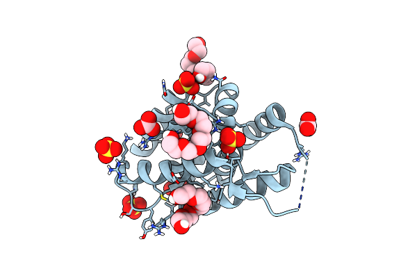 |
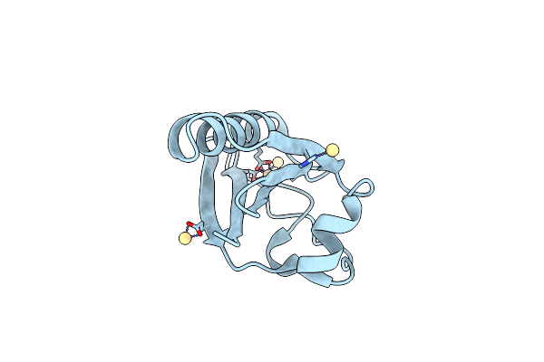
Organism: Bacillus subtilis
Method: X-RAY DIFFRACTION Resolution:1.15 Å Release Date: 2024-05-01 Classification: CHAPERONE Ligands: 1PE, CO3, GOL, SO4 |
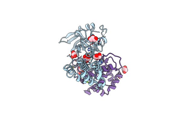 |
Organism: Bacillus subtilis
Method: X-RAY DIFFRACTION Resolution:2.09 Å Release Date: 2023-12-27 Classification: UNKNOWN FUNCTION Ligands: GOL, MG |
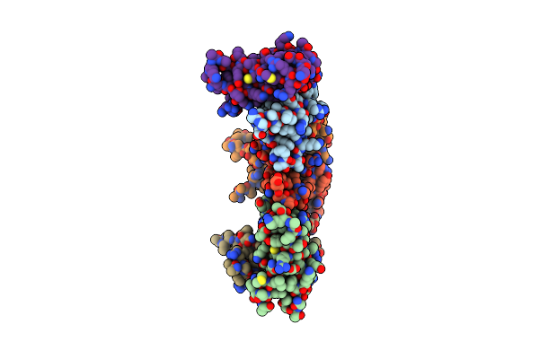 |
Organism: Bacillus subtilis
Method: X-RAY DIFFRACTION Resolution:2.38 Å Release Date: 2023-08-23 Classification: DNA BINDING PROTEIN |
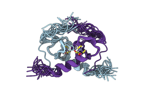 |
Organism: Bacillus subtilis
Method: SOLUTION NMR Release Date: 2018-02-28 Classification: TRANSCRIPTION Ligands: ZN |
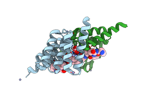 |
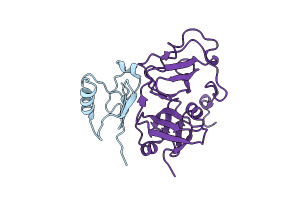
Structure Of The Tpr Domain Of Sgt2 In Complex With Yeast Ssa1 Peptide Fragment
Organism: Saccharomyces cerevisiae
Method: X-RAY DIFFRACTION Resolution:2.00 Å Release Date: 2017-10-25 Classification: CHAPERONE Ligands: ZN |
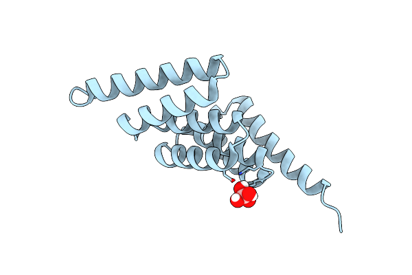 |
Organism: Saccharomyces cerevisiae
Method: X-RAY DIFFRACTION Resolution:1.55 Å Release Date: 2017-10-25 Classification: CHAPERONE Ligands: BO4 |
 |
Organism: Bacillus subtilis
Method: SOLUTION NMR Release Date: 2017-06-21 Classification: TRANSCRIPTION Ligands: ZN |
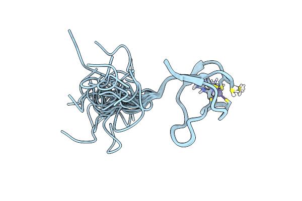 |
Organism: Homo sapiens
Method: SOLUTION NMR Release Date: 2016-05-25 Classification: LIGASE Ligands: ZN |
 |
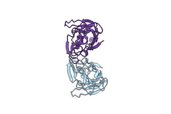
Solution Structure Of Rnf126 N-Terminal Zinc Finger Domain In Complex With Bag6 Ubiquitin-Like Domain
Organism: Homo sapiens
Method: SOLUTION NMR Release Date: 2016-05-25 Classification: LIGASE Ligands: ZN |
 |
 |
Organism: Saccharomyces cerevisiae
Method: SOLUTION NMR Release Date: 2013-01-16 Classification: CHAPERONE |
 |
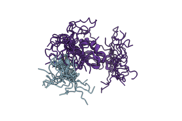
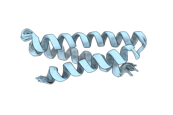
Structure Of The Complex Between The N-Terminal Dimerisation Domain Of Sgt2 And The Ubl Domain Of Get5
Organism: Saccharomyces cerevisiae
Method: SOLUTION NMR Release Date: 2013-01-16 Classification: CHAPERONE |
 |
Organism: Saccharomyces cerevisiae
Method: X-RAY DIFFRACTION Resolution:1.78 Å Release Date: 2012-11-14 Classification: PROTEIN BINDING Ligands: SO4 |
 |
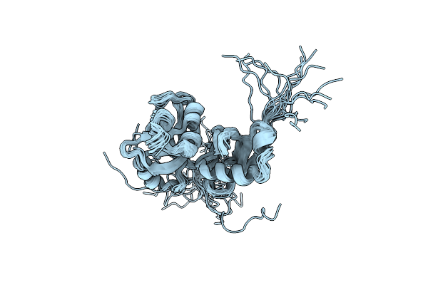
Solution Structure Of The Alpha Subdomain Of The Major Non-Repeat Unit Of Fap1 Fimbriae Of Streptococcus Parasanguis
Organism: Streptococcus parasanguinis
Method: SOLUTION NMR Release Date: 2010-07-21 Classification: STRUCTURAL PROTEIN |
 |
Ph-Induced Modulation Of Streptococcus Parasanguinis Adhesion By Fap1 Fimbriae
Organism: Streptococcus parasanguinis
Method: X-RAY DIFFRACTION Resolution:2.90 Å Release Date: 2010-07-07 Classification: CELL ADHESION |
 |
Organism: Mus musculus
Method: SOLUTION NMR Release Date: 2007-05-08 Classification: TRANSPORT PROTEIN |
 |
Ufd1 Exhibits The Aaa-Atpase Fold With Two Distinct Ubiquitin Interaction Sites
Organism: Saccharomyces cerevisiae
Method: SOLUTION NMR Release Date: 2005-07-26 Classification: PROTEIN TURNOVER |
 |
Organism: Homo sapiens
Method: X-RAY DIFFRACTION Resolution:1.65 Å Release Date: 2002-01-04 Classification: STRUCTURAL PROTEIN Ligands: CD |