
Search Count: 58
 |
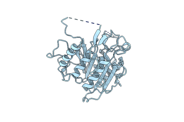
Organism: Trypanosoma brucei
Method: X-RAY DIFFRACTION Resolution:1.40 Å Release Date: 2012-05-02 Classification: HYDROLASE |
 |
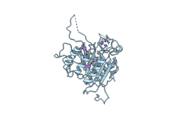
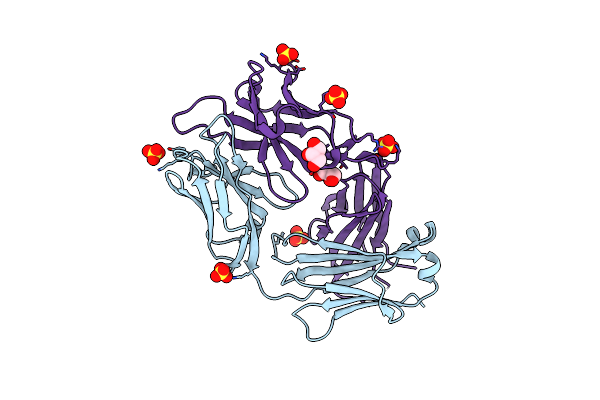
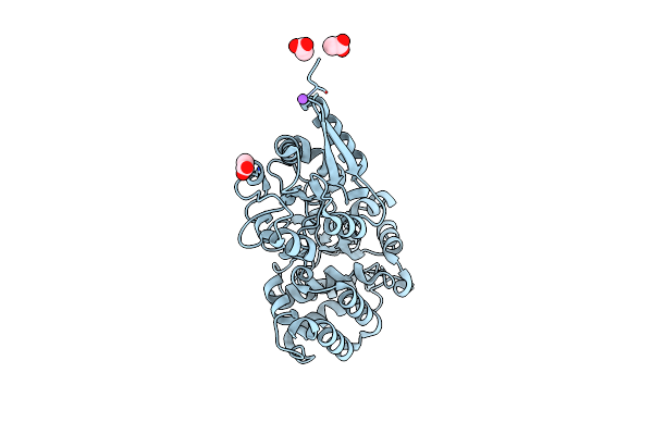
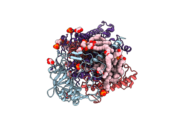
The Structure Of Metacaspase 2 From T. Brucei Determined In The Presence Of Samarium
Organism: Trypanosoma brucei
Method: X-RAY DIFFRACTION Resolution:2.10 Å Release Date: 2012-05-02 Classification: HYDROLASE Ligands: SM |
 |
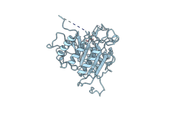
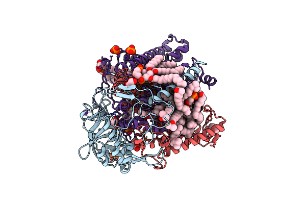
Organism: Trypanosoma brucei brucei
Method: X-RAY DIFFRACTION Resolution:1.60 Å Release Date: 2012-05-02 Classification: HYDROLASE |
 |
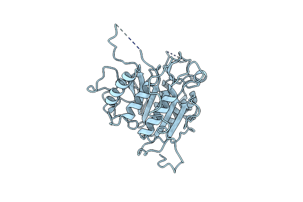
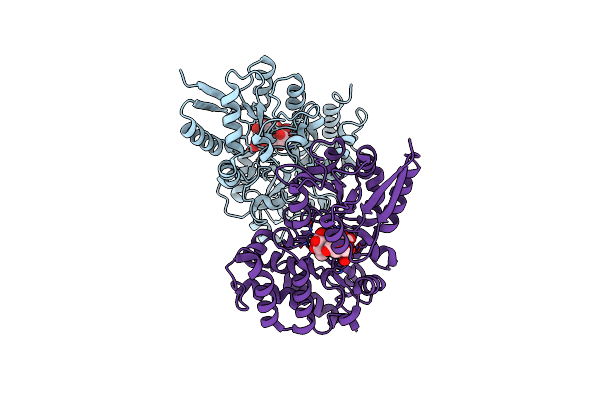
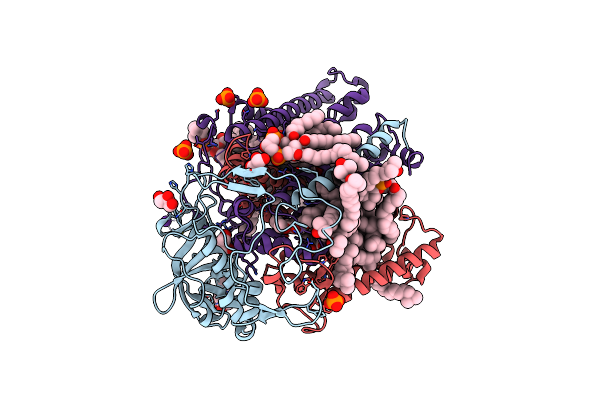
The Structure Of Metacaspase 2 From T. Brucei Determined In The Presence Of Calcium Chloride
Organism: Trypanosoma brucei
Method: X-RAY DIFFRACTION Resolution:1.50 Å Release Date: 2012-05-02 Classification: HYDROLASE |
 |
Crystal Structure Of The Reaction Centre From Blastochloris Viridis Strain Dsm 133 (Atcc 19567) Substrain-08
Organism: Blastochloris viridis
Method: X-RAY DIFFRACTION Resolution:1.95 Å Release Date: 2011-11-23 Classification: ELECTRON TRANSPORT Ligands: HEC, LDA, DGA, SO4, HTO, GOL, BCB, BPB, UQ9, FE2, MQ9, NS5, HTH |
 |
Crystal Structure Of The Reaction Centre From Blastochloris Viridis Strain Dsm 133 (Atcc 19567) Substrain-94
Organism: Blastochloris viridis
Method: X-RAY DIFFRACTION Resolution:1.92 Å Release Date: 2011-11-23 Classification: ELECTRON TRANSPORT Ligands: HEC, LDA, DGA, SO4, HTO, GOL, BCB, BPB, UQ9, MQ9, FE2, NS5 |
 |
Organism: Human adenovirus 5
Method: X-RAY DIFFRACTION Resolution:1.57 Å Release Date: 2011-11-02 Classification: VIRAL PROTEIN |
 |
Crystal Structure Of The Beta Domain Of The Bordetella Autotransporter Brka
Organism: Bordetella pertussis
Method: X-RAY DIFFRACTION Resolution:3.00 Å Release Date: 2011-04-13 Classification: MEMBRANE PROTEIN/PROTEIN TRANSPORT |
 |
Organism: Rhodobacter sphaeroides
Method: X-RAY DIFFRACTION Resolution:2.01 Å Release Date: 2010-12-01 Classification: MEMBRANE PROTEIN Ligands: BCL, BPH, U10, UQ1, PO4, DIO, GOL, HT3, HTO, LDA, FE, SPO, K, CL, CDL |
 |
Organism: Leishmania major, Actinobacteria
Method: X-RAY DIFFRACTION Resolution:1.65 Å Release Date: 2010-10-06 Classification: HYDROLASE/INHIBITOR Ligands: GOL, PGR, PGO, PO4, CL, NA |
 |
Organism: Mus musculus
Method: X-RAY DIFFRACTION Resolution:1.95 Å Release Date: 2010-07-21 Classification: IMMUNE SYSTEM Ligands: SO4, CL, GOL |
 |
Organism: Enterococcus faecalis
Method: X-RAY DIFFRACTION Resolution:1.69 Å Release Date: 2010-06-16 Classification: STRUCTURAL PROTEIN Ligands: CL |
 |
Crystal Structure Of Rafe From Streptococcus Pneumoniae Complexed With Raffinose
Organism: Streptococcus pneumoniae
Method: X-RAY DIFFRACTION Resolution:2.80 Å Release Date: 2007-08-07 Classification: SUGAR BINDING PROTEIN Ligands: CL |
 |
Organism: Streptococcus pneumoniae
Method: X-RAY DIFFRACTION Resolution:1.40 Å Release Date: 2007-07-31 Classification: SUGAR BINDING PROTEIN |
 |
Atomic Resolution Structure Of Apo-Form Of Rafe From Streptococcus Pneumoniae
Organism: Streptococcus pneumoniae
Method: X-RAY DIFFRACTION Resolution:1.04 Å Release Date: 2007-06-05 Classification: TRANSPORT PROTEIN Ligands: CL, NA, TRS |
 |
Crystal Structure Of Selenomethionine-Labelled Rafe From Streptococcus Pneumoniae
Organism: Streptococcus pneumoniae
Method: X-RAY DIFFRACTION Resolution:2.90 Å Release Date: 2007-06-05 Classification: SUGAR BINDING PROTEIN Ligands: CL |
 |
Reaction Centre From Rhodobacter Sphaeroides Strain R-26.1 Complexed With Brominated Phosphatidylcholine
Organism: Rhodobacter sphaeroides
Method: X-RAY DIFFRACTION Resolution:2.70 Å Release Date: 2007-03-27 Classification: PHOTOSYNTHESIS/MEMBRANE PROTEIN Ligands: PO4, BCL, BPH, U10, HTO, LDA, GOL, FE, CDL, PC9, K |
 |
Reaction Centre From Rhodobacter Sphaeroides Strain R-26.1 Complexed With Tetrabrominated Phosphatidylcholine
Organism: Rhodobacter sphaeroides
Method: X-RAY DIFFRACTION Resolution:2.45 Å Release Date: 2007-03-27 Classification: PHOTOSYNTHESIS/MEMBRANE PROTEIN Ligands: PO4, CL, BCL, BPH, U10, FE, CDL, PCK, LDA, GOL, K, PC7 |
 |
Reaction Centre From Rhodobacter Sphaeroides Strain R-26.1 Complexed With Dibrominated Phosphatidylcholine
Organism: Rhodobacter sphaeroides
Method: X-RAY DIFFRACTION Resolution:2.55 Å Release Date: 2007-03-27 Classification: PHOTOSYNTHESIS/MEMBRANE PROTEIN Ligands: PO4, BCL, BPH, U10, PC9, LDA, FE, CDL, K, HTO, PC7, GOL |
 |
Reaction Centre From Rhodobacter Sphaeroides Strain R-26.1 Complexed With Dibrominated Phosphatidylglycerol
Organism: Rhodobacter sphaeroides
Method: X-RAY DIFFRACTION Resolution:2.50 Å Release Date: 2007-03-27 Classification: PHOTOSYNTHESIS/MEMBRANE PROTEIN Ligands: BCL, BPH, U10, GOL, FE, CL, PO4, CDL, PGK, LDA, K, PGT |