
Search Count: 67
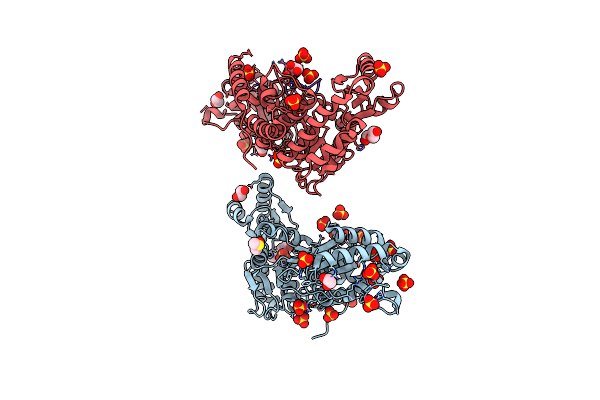 |
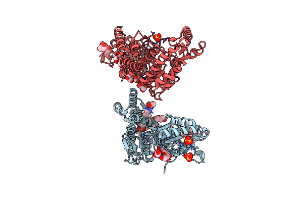
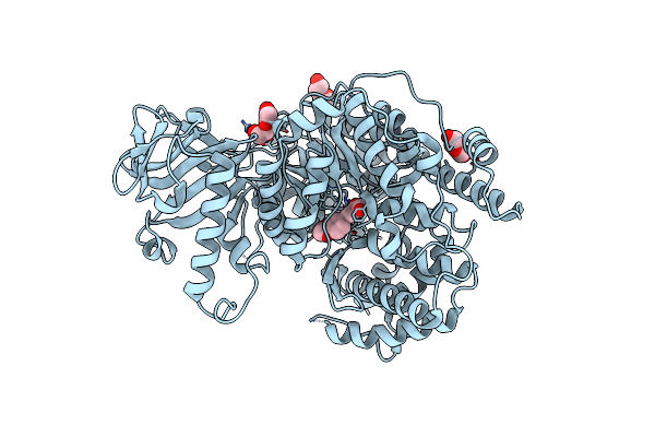
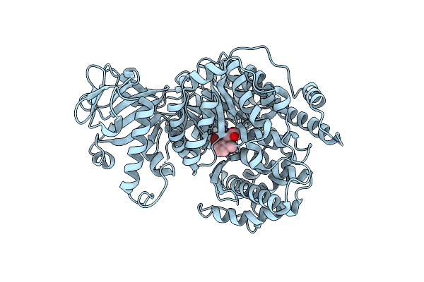
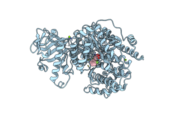
Mycobacterium Tuberculosis Pks13 Acyltransferase Serine Converted To Beta-Lactam Form By Cec215 Via Sufex Reaction
Organism: Mycobacterium tuberculosis h37rv
Method: X-RAY DIFFRACTION Release Date: 2025-05-21 Classification: ANTIBIOTIC Ligands: SO4, CL, PG4, EDO, DMS, GOL, PEG |
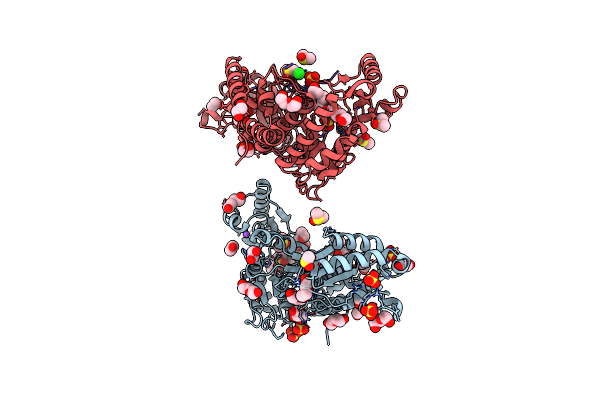 |
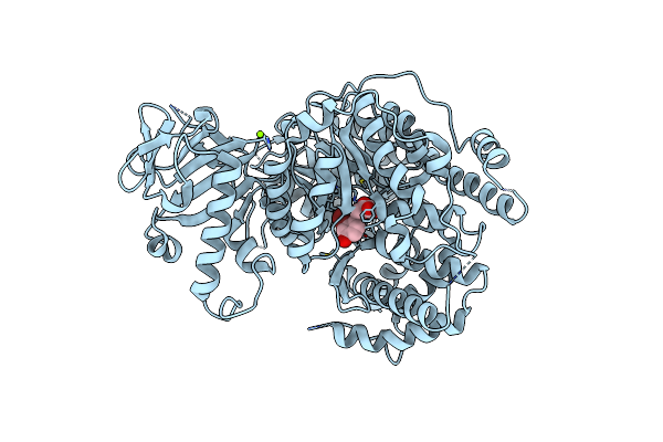
Organism: Mycobacterium tuberculosis (strain atcc 25618 / h37rv)
Method: X-RAY DIFFRACTION Release Date: 2025-05-21 Classification: ANTIBIOTIC Ligands: DMS, SO4, PE5, EDO, PEG, GOL, CL, NA, P33 |
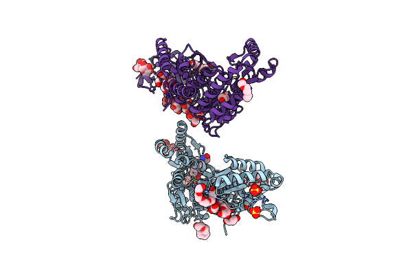 |
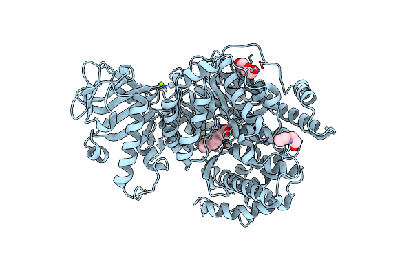
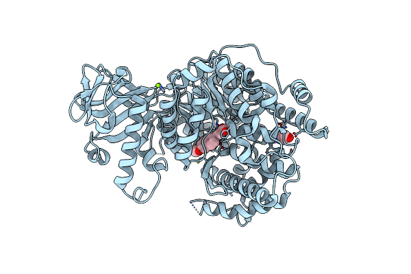
M. Tuberculosis Pks13 Acyltransferase (At) Domain In Complex With Sufex Inhibitor Cec215
Organism: Mycobacterium tuberculosis
Method: X-RAY DIFFRACTION Release Date: 2025-05-07 Classification: TRANSFERASE/TRANSFERASE INHIBITOR Ligands: 1PE, SO4, A1ATV |
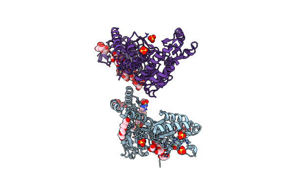 |
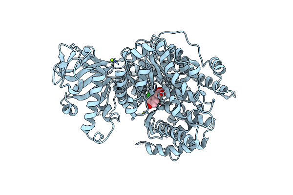
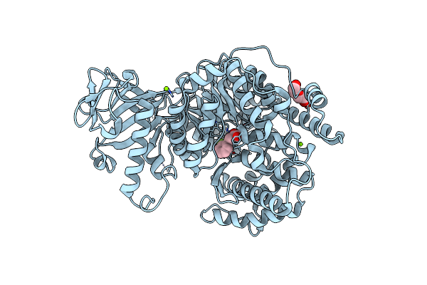
M. Tuberculosis Pks13 Acyltransferase (At) Domain In Complex With Sufex Inhibitor Cmx410
Organism: Mycobacterium tuberculosis (strain atcc 25618 / h37rv)
Method: X-RAY DIFFRACTION Release Date: 2025-05-07 Classification: TRANSFERASE/TRANSFERSE INHIBITOR Ligands: 1PE, SO4, A1ATW |
 |
Organism: Mycobacterium tuberculosis
Method: X-RAY DIFFRACTION Release Date: 2025-05-07 Classification: TRANSFERASE Ligands: 1PE, SO4 |
 |
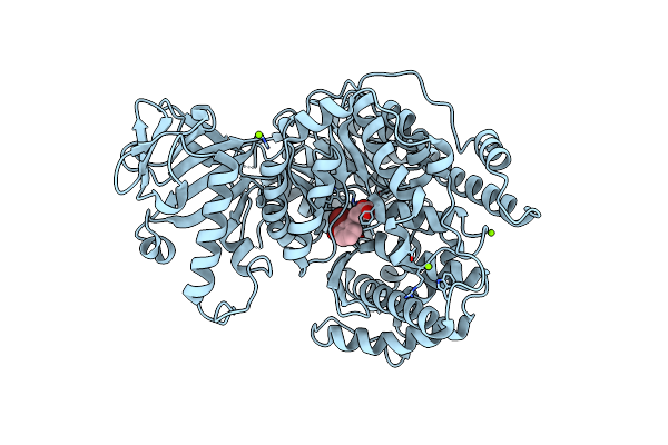
M. Tuberculosis Pks13 Acyltransferase (At) Domain In Complex With Sufex Inhibitor Cmx410 - Reaction Product
Organism: Mycobacterium tuberculosis (strain atcc 25618 / h37rv)
Method: X-RAY DIFFRACTION Release Date: 2025-05-07 Classification: TRANSFERASE Ligands: SO4, 1PE, A1AVL |
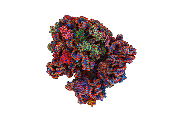 |
Cryo-Em Structure Of The E.Coli 70S Ribosome In Complex With The Antibiotic Myxovalargin B.
Organism: Myxococcus fulvus, Escherichia coli k-12
Method: ELECTRON MICROSCOPY Release Date: 2023-01-25 Classification: RIBOSOME Ligands: MG, ZN, FME, SPD |
 |
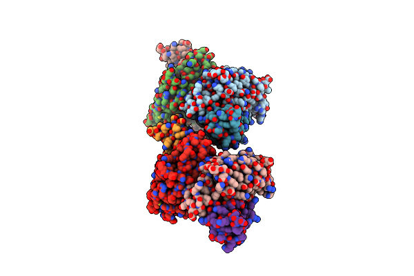
Cryo-Em Structure Of The E.Coli 50S Ribosomal Subunit In Complex With The Antibiotic Myxovalargin A.
Organism: Myxococcus fulvus, Escherichia coli k-12
Method: ELECTRON MICROSCOPY Release Date: 2023-01-18 Classification: RIBOSOME Ligands: MG, ZN |
 |
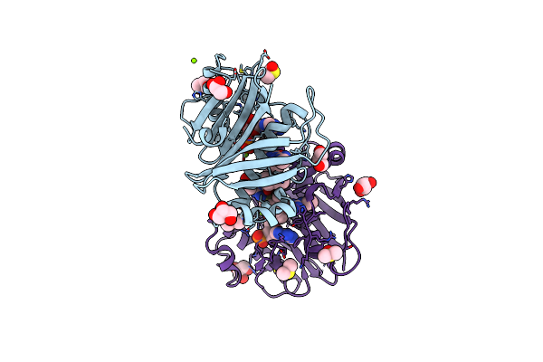
Mycobacterium Tuberculosis 3-Hydroxyl-Acp Dehydratase Hadab In Complex With 1,3-Diarylpyrazolyl-Acylsulfonamide Inhibitor
Organism: Mycobacterium tuberculosis
Method: X-RAY DIFFRACTION Resolution:2.40 Å Release Date: 2022-11-16 Classification: LYASE/INHIBITOR Ligands: CI7, EDO, PEG, GOL |
 |
Organism: Mycobacterium tuberculosis
Method: X-RAY DIFFRACTION Resolution:1.76 Å Release Date: 2019-02-06 Classification: TRANSFERASE/INHIBITOR Ligands: COA, FD7, MG, GOL, DMS |
 |
Crystal Structure Of Mycobacterium Tuberculosis Malate Synthase In Complex With Dioxine-Phenyldiketoacid
Organism: Mycobacterium tuberculosis
Method: X-RAY DIFFRACTION Resolution:2.10 Å Release Date: 2018-09-05 Classification: TRANSFERASE/TRANSFERASE inhibitor Ligands: MG, BXS, PEG |
 |
Crystal Structure Of Mycobacterium Tuberculosis Malate Synthase In Complex With 2-Naphthyldiketoacid
Organism: Mycobacterium tuberculosis
Method: X-RAY DIFFRACTION Resolution:1.80 Å Release Date: 2018-09-05 Classification: TRANSFERASE/TRANSFERASE inhibitor Ligands: MG, C0V, PEG |
 |
Crystal Structure Of Mycobacterium Tuberculosis Malate Synthase In Complex With 2-Cl-4-Oh-Phenyldiketoacid
Organism: Mycobacterium tuberculosis
Method: X-RAY DIFFRACTION Resolution:2.50 Å Release Date: 2018-09-05 Classification: TRANSFERASE/TRANSFERASE inhibitor Ligands: MG, D1Y |
 |
Crystal Structure Of Mycobacterium Tuberculosis Malate Synthase In Complex With 2-Br-3-Oh-Phenyldiketoacid
Organism: Mycobacterium tuberculosis
Method: X-RAY DIFFRACTION Resolution:1.58 Å Release Date: 2018-09-05 Classification: STRUCTURAL GENOMICS,Transferase Ligands: MG, E9S |
 |
Crystal Structure Of Mycobacterium Tuberculosis Malate Synthase In Complex With 2-Br-6-Me-Phenyldiketoacid
Organism: Mycobacterium tuberculosis
Method: X-RAY DIFFRACTION Resolution:2.60 Å Release Date: 2018-09-05 Classification: TRANSFERASE/TRANSFERASE inhibitor Ligands: MG, EHV |
 |
Crystal Structure Of Mycobacterium Tuberculosis Malate Synthase In Complex With 2-Br-4-Oh-Phenyldiketoacid
Organism: Mycobacterium tuberculosis
Method: X-RAY DIFFRACTION Resolution:2.30 Å Release Date: 2018-09-05 Classification: TRANSFERASE/TRANSFERASE inhibitor Ligands: MG, ENG |
 |
Crystal Structure Of Mycobacterium Tuberculosis Malate Synthase In Complex With Methoxynaphthyldiketoacid
Organism: Mycobacterium tuberculosis
Method: X-RAY DIFFRACTION Resolution:2.13 Å Release Date: 2018-09-05 Classification: TRANSFERASE/TRANSFERASE inhibitor Ligands: ENY, MG, PEG |
 |
Crystal Structure Of Mycobacterium Tuberculosis Malate Synthase In Complex With 2-F-Phenyldiketoacid
Organism: Mycobacterium tuberculosis
Method: X-RAY DIFFRACTION Resolution:1.64 Å Release Date: 2018-09-05 Classification: TRANSFERASE Ligands: MG, EQA, PEG |
 |
Crystal Structure Of Mycobacterium Tuberculosis Malate Synthase In Complex With 2,6-F-Phenyldiketoacid
Organism: Mycobacterium tuberculosis
Method: X-RAY DIFFRACTION Resolution:1.56 Å Release Date: 2018-09-05 Classification: LYASE/LYASE inhibitor Ligands: MG, GXG |
 |
Crystal Structure Of Mycobacterium Tuberculosis Malate Synthase In Complex With 2,6-Cl-Phenyldiketoacid
Organism: Mycobacterium tuberculosis
Method: X-RAY DIFFRACTION Resolution:1.80 Å Release Date: 2018-09-05 Classification: LYASE/LYASE inhibitor Ligands: MG, GXD |