
Search Count: 52
 |
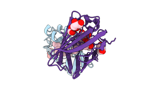
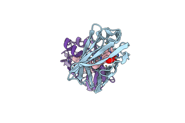
Q108K:K40L:T51V:T53S:R58W:Y19W:L117E Mutant Of Hcrbpii Bound To Fluorophore Td-1V-3
Organism: Homo sapiens
Method: X-RAY DIFFRACTION Release Date: 2025-10-29 Classification: RETINOL BINDING PROTEIN Ligands: A1CKW, ACT |
 |
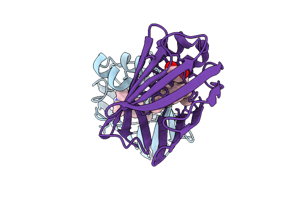
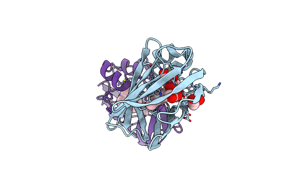
Q108K:K40L:T51V:T53S:R58W:Y19W:L117E Mutant Of Hcrbpii Bound To Fluorophore Td-1V-9
Organism: Homo sapiens
Method: X-RAY DIFFRACTION Release Date: 2025-10-29 Classification: RETINOL BINDING PROTEIN Ligands: ACT, A1CK1 |
 |
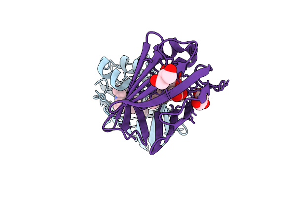
Q108K:K40L:T51V:T53S:R58W:Y19W:L117E Mutant Of Hcrbpii Bound To Fluorophore Td-1V-2
Organism: Homo sapiens
Method: X-RAY DIFFRACTION Release Date: 2025-10-01 Classification: RETINOL BINDING PROTEIN Ligands: A1CKV, ACT |
 |
Q108K:K40L:T51V:T53S:R58W:Y19W:L117E Mutant Of Hcrbpii Bound To Fluorophore Td-1V-1
Organism: Homo sapiens
Method: X-RAY DIFFRACTION Release Date: 2025-10-01 Classification: RETINOL BINDING PROTEIN Ligands: A1CKX, ACT |
 |
Q108K:K40L:T51V:T53S:R58W:Y19W:L117E Mutant Of Hcrbpii Bound To Fluorophore Td-1V-6
Organism: Homo sapiens
Method: X-RAY DIFFRACTION Release Date: 2025-10-01 Classification: RETINOL BINDING PROTEIN Ligands: A1CKY, ACT, GOL |
 |
Q108K:K40L:T51V:T53S:R58W:Y19W:L117E Mutant Of Hcrbpii Bound To Fluorophore Td-1V-8
Organism: Homo sapiens
Method: X-RAY DIFFRACTION Release Date: 2025-10-01 Classification: RETINOL BINDING PROTEIN Ligands: A1CKZ, ACT |
 |
Q108K:K40L:T51V:T53S:R58W:Y19W:L117E Mutant Of Hcrbpii Bound To Fluorophore Td-1V-10
Organism: Homo sapiens
Method: X-RAY DIFFRACTION Release Date: 2025-10-01 Classification: RETINOL BINDING PROTEIN Ligands: A1CK0, ACT |
 |
Q108K:K40L:T51V:T53C:R58Y:Y19W:Q38L:Q4A:T29L Mutant Of Hcrbpii Bound To Fr1V In The Dark At Ph 6.0
Organism: Homo sapiens
Method: X-RAY DIFFRACTION Release Date: 2025-09-03 Classification: RETINOL BINDING PROTEIN Ligands: ZFJ, ACT, GOL, MLI |
 |
Q108K:K40L:T51V:T53C:R58A:Y19W:Q38L:Q4A:T29L Mutant Of Hcrbpii Bound To Fr1V And Irradiated With Uv Light At Ph 3.0
Organism: Homo sapiens
Method: X-RAY DIFFRACTION Release Date: 2025-09-03 Classification: RETINOL BINDING PROTEIN Ligands: ACT, GOL, ZFJ |
 |
Q108K:K40L:T51V:T53C:R58A:Y19W:Q38L:Q4A:T29L Mutant Of Hcrbpii Bound To Fr1V In The Dark At Ph 3.0
Organism: Homo sapiens
Method: X-RAY DIFFRACTION Release Date: 2025-09-03 Classification: RETINOL BINDING PROTEIN Ligands: ZFJ, EDO |
 |
Q108K:K40L:T51V:T53C:R58Y:Y19W:Q38L:Q4A:T29L Mutant Of Hcrbpii In Complex With Fr1V In The Dark State At Ph 3.0
Organism: Homo sapiens
Method: X-RAY DIFFRACTION Release Date: 2025-07-16 Classification: RETINOL BINDING PROTEIN Ligands: ACT, GOL, ZFJ |
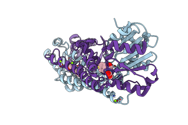 |
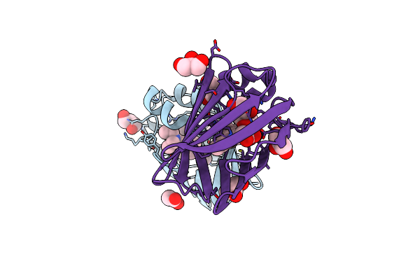
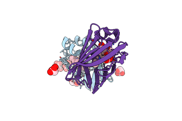
Fphe, Staphylococcus Aureus Fluorophosphonate-Binding Serine Hydrolases E, Oxadiazolone-Peptide Bound
Organism: Staphylococcus aureus subsp. aureus usa300
Method: X-RAY DIFFRACTION Resolution:1.54 Å Release Date: 2025-03-12 Classification: HYDROLASE Ligands: A1BA1, MG |
 |
Q108K:K40L:T51V:T53C:R58W:Y19W:Q38L:Q4A:T29L Mutant Of Hcrbpii Bound To Fr1V Fluorophore In The Dark State At Ph 4
Organism: Homo sapiens
Method: X-RAY DIFFRACTION Resolution:1.20 Å Release Date: 2025-02-12 Classification: RETINOL BINDING PROTEIN Ligands: ZFJ, ACY |
 |
Q108K:K40L:T51V:T53C:R58W:Y19W:Q38L:Q4A:T29L Mutant Of Hcrbpii Bound To Fr1V And Irradiated With Uv Light At Ph 4
Organism: Homo sapiens
Method: X-RAY DIFFRACTION Resolution:1.30 Å Release Date: 2025-02-12 Classification: RETINOL BINDING PROTEIN Ligands: ZFJ, GOL, ACY |
 |
Q108K:K40L:T51V:T53C:R58Y:Y19W:Q38L:Q4A:T29L Mutant Of Hcrbpii Bound To Fr1V And Irradiated With Uv Light At Ph 3.0
Organism: Homo sapiens
Method: X-RAY DIFFRACTION Resolution:1.60 Å Release Date: 2025-02-12 Classification: RETINOL BINDING PROTEIN Ligands: ZFJ, ACY, GOL |
 |
Q108K:K40L:T51V:T53C:R58Y:Y19W:Q38L:Q4A:T29L Mutant Of Hcrbpii Bound To Fr1V And Irradiated With Uv Light At Ph 7.6
Organism: Homo sapiens
Method: X-RAY DIFFRACTION Resolution:1.52 Å Release Date: 2025-02-12 Classification: RETINOL BINDING PROTEIN Ligands: ZFJ, ACY, MLA, GOL |
 |
Crystal Structure Of Bama In Complex With The Ptb2 Open-State Inhibitor (Anisotropic Data Set)
Organism: Escherichia coli, Synthetic construct
Method: X-RAY DIFFRACTION Resolution:2.75 Å Release Date: 2025-01-15 Classification: MEMBRANE PROTEIN |
 |
Organism: Escherichia coli
Method: ELECTRON MICROSCOPY Release Date: 2024-12-25 Classification: MEMBRANE PROTEIN |
 |
Cryoem Structure Of Bam In Complex With The Ptb1 Closed-State Inhibitor (In Ddm Detergent)
Organism: Escherichia coli, Synthetic construct
Method: ELECTRON MICROSCOPY Release Date: 2024-12-25 Classification: MEMBRANE PROTEIN Ligands: ZN |
 |
Organism: Escherichia coli
Method: ELECTRON MICROSCOPY Release Date: 2024-12-25 Classification: MEMBRANE PROTEIN |