
Search Count: 308
 |
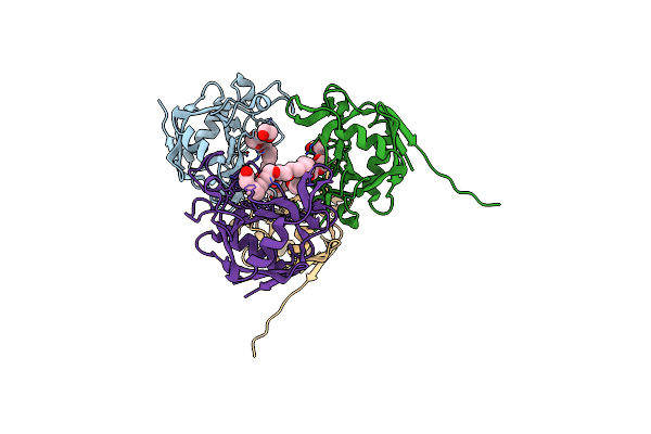
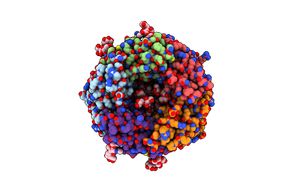
Organism: Hepatitis e virus (strain pakistan), Vicugna pacos
Method: X-RAY DIFFRACTION Release Date: 2025-09-03 Classification: VIRAL PROTEIN/IMMUNE SYSTEM |
 |
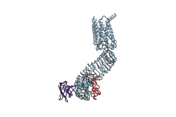
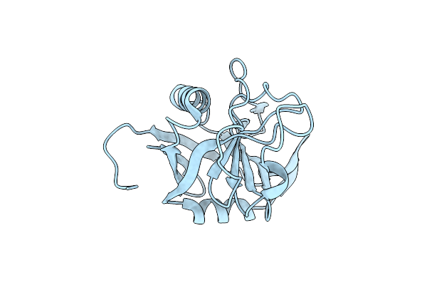
Organism: Homo sapiens, Camelus bactrianus
Method: ELECTRON MICROSCOPY Release Date: 2025-01-15 Classification: MEMBRANE PROTEIN |
 |
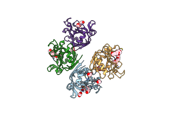
Organism: Camelus bactrianus, Homo sapiens
Method: ELECTRON MICROSCOPY Release Date: 2025-01-15 Classification: MEMBRANE PROTEIN |
 |
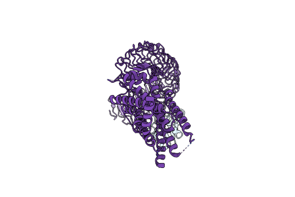
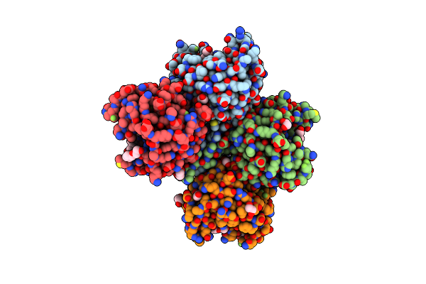
Crystal Structure Of Maltose Binding Protein (Apo), Mutant Trp10 To 4-Cyanotryptophan
Organism: Escherichia coli
Method: X-RAY DIFFRACTION Resolution:1.48 Å Release Date: 2024-12-18 Classification: SUGAR BINDING PROTEIN |
 |
Organism: Escherichia coli
Method: X-RAY DIFFRACTION Resolution:1.58 Å Release Date: 2024-12-18 Classification: SUGAR BINDING PROTEIN Ligands: PEG, EDO, PGE, NA, CD |
 |
E. Coli Peptidyl-Prolyl Cis-Trans Isomerase Containing (2S,3S)-4-Fluorovaline
Organism: Escherichia coli
Method: X-RAY DIFFRACTION Resolution:1.30 Å Release Date: 2024-10-09 Classification: ISOMERASE Ligands: 1PE, EDO, PGE, ACT, PEG, XPE |
 |
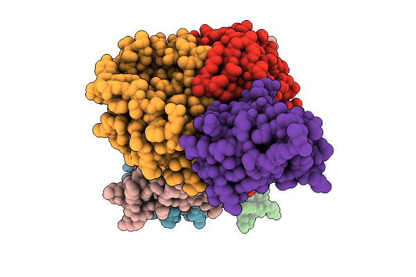
Cryoem Structure Of Human Full-Length Alpha1Beta3Gamma2L Gaba(A)R In Complex With Gaba And Puerarin
Organism: Homo sapiens
Method: ELECTRON MICROSCOPY Release Date: 2024-09-18 Classification: MEMBRANE PROTEIN Ligands: PGW, D10, HEX, PT5, R16, PLM, CL, ABU, NAG, A1H6W, PX2 |
 |
Solution Structure Of Toxin, U-Rdtx-Pp19, From Assassin Bug Pristhesancus Plagipennis
Organism: Pristhesancus plagipennis
Method: SOLUTION NMR Release Date: 2024-05-29 Classification: TOXIN |
 |
E. Coli Peptidyl-Prolyl Cis-Trans Isomerase Containing Delta1-Monofluoro-Leucines
Organism: Escherichia coli
Method: X-RAY DIFFRACTION Resolution:1.22 Å Release Date: 2024-05-22 Classification: ISOMERASE |
 |
E. Coli Peptidyl-Prolyl Cis-Trans Isomerase Containing Delta2-Monofluoro-Leucines
Organism: Escherichia coli
Method: X-RAY DIFFRACTION Resolution:1.80 Å Release Date: 2024-05-22 Classification: ISOMERASE Ligands: EDO, PGE, PEG, 1PE, XPE |
 |
Organism: Escherichia coli
Method: X-RAY DIFFRACTION Resolution:1.65 Å Release Date: 2024-05-22 Classification: ISOMERASE Ligands: PEG, PGE, EDO, PG4, 2PE |
 |
Structure Of Human Hypoxanthine Guanine Phosphoribzosyltransferase In Complex With [2S,4R]-4-Guanin-9-Yl-2-(2-Phosphonoethoxymethyl)-1-N-(3-Phosphonopropionyl)Pyrrolidine
Organism: Homo sapiens
Method: X-RAY DIFFRACTION Resolution:2.27 Å Release Date: 2024-05-08 Classification: TRANSFERASE Ligands: JEI, MG |
 |
Structure Of Human Hypoxanthine Guanine Phosphoribzosyltransferase In Complex With [2S,4R] 4-Guanin-9-Yl-2-Hydroxymethyl-1-N-(3-Phosphonopropionyl)Pyrrolidine
Organism: Homo sapiens
Method: X-RAY DIFFRACTION Resolution:2.50 Å Release Date: 2024-05-08 Classification: TRANSFERASE Ligands: JG6, MG |
 |
Crystal Structure Of Trypanosome Brucei Hypoxanthine Guanine Phosphopribosyltransferase In Complex With [2S,4R]-4-Guanin-9-Yl-2-(2- Phosphonoethoxymethyl)-1-N-(3-Phosphonopropionyl)Pyrrolidine
Organism: Trypanosoma brucei
Method: X-RAY DIFFRACTION Resolution:2.46 Å Release Date: 2024-05-08 Classification: TRANSFERASE Ligands: JEI |
 |
Crystal Structure Of T. Brucei Hypoxanthine Guanine Phosphoribosyltransferase In Complex With [2S,4S]-4-Guanin-9-Yl-2-(2-Phosphonoethoxymethyl)-1-N-(3-Phosphonopropionyl)Pyrrolidine
Organism: Trypanosoma brucei
Method: X-RAY DIFFRACTION Resolution:2.20 Å Release Date: 2024-05-08 Classification: TRANSFERASE Ligands: MG, KFF |
 |
Organism: Mus musculus
Method: ELECTRON MICROSCOPY Release Date: 2024-02-14 Classification: VIRUS LIKE PARTICLE |
 |
Organism: Mus musculus
Method: ELECTRON MICROSCOPY Release Date: 2024-02-14 Classification: VIRUS LIKE PARTICLE |
 |
Organism: Mus musculus
Method: ELECTRON MICROSCOPY Release Date: 2024-02-14 Classification: VIRUS LIKE PARTICLE |
 |
Organism: Mus musculus
Method: ELECTRON MICROSCOPY Release Date: 2024-02-14 Classification: VIRUS LIKE PARTICLE |
 |
Organism: Acinetobacter baumannii 1295743
Method: X-RAY DIFFRACTION Resolution:3.03 Å Release Date: 2023-10-11 Classification: HYDROLASE |