
Search Count: 83
 |
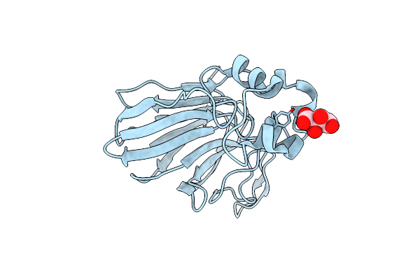
Organism: Homo sapiens
Method: X-RAY DIFFRACTION Release Date: 2025-10-01 Classification: ISOMERASE Ligands: SO4 |
 |
 |
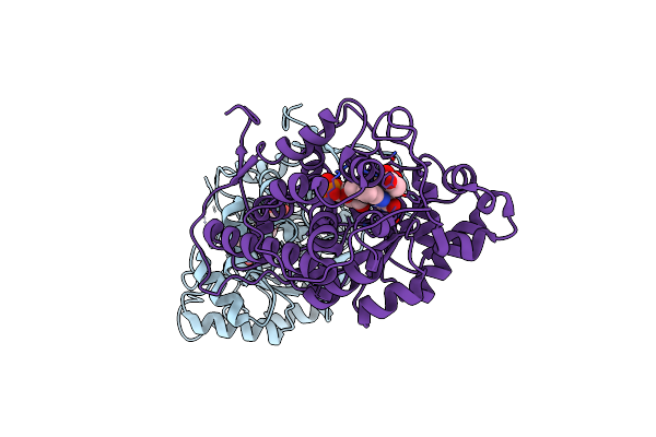
Organism: Staphylococcus aureus
Method: X-RAY DIFFRACTION Release Date: 2025-04-30 Classification: HYDROLASE Ligands: OCA, PPI, GOL, A1L60, FMT, ZN, CA, 6NA, CL, BUA, 11A |
 |
Neutron Structure Of Cellulase Cel6A From Phanerochaete Chrysosporium At Room Temperature
Organism: Phanerodontia chrysosporium
Method: X-RAY DIFFRACTION, NEUTRON DIFFRACTION Resolution:1.36 Å, 1.86 Å Release Date: 2025-03-12 Classification: HYDROLASE |
 |
Neutron Structure Of Cellulase Cel6A From Phanerochaete Chrysosporium At Room Temperature, Enzyme-Product Complex
Organism: Phanerodontia chrysosporium
Method: X-RAY DIFFRACTION, NEUTRON DIFFRACTION Resolution:1.40 Å, 1.8 Å Release Date: 2025-03-12 Classification: HYDROLASE Ligands: BGC, NA |
 |
Neutron Structure Of Cellulase Cel6A From Phanerochaete Chrysosporium At Room Temperature, Low-D2O-Solvent
Organism: Phanerodontia chrysosporium
Method: X-RAY DIFFRACTION, NEUTRON DIFFRACTION Resolution:1.40 Å, 2.15 Å Release Date: 2025-03-12 Classification: HYDROLASE |
 |
Neutron Structure Of Cellulase Cel6A From Phanerochaete Chrysosporium At Room Temperature, Enzyme-Product Complex, H2O Solvent
Organism: Phanerodontia chrysosporium
Method: X-RAY DIFFRACTION, NEUTRON DIFFRACTION Resolution:1.80 Å, 2.59 Å Release Date: 2025-03-12 Classification: HYDROLASE Ligands: BGC, NA |
 |
High-Resolution X-Ray Structure Of Cellulase Cel6A From Phanerochaete Chrysosporium At Cryogenic Temperature
Organism: Phanerodontia chrysosporium
Method: X-RAY DIFFRACTION Resolution:0.80 Å Release Date: 2025-03-12 Classification: HYDROLASE Ligands: MPD, ACT |
 |
High-Resolution X-Ray Structure Of Cellulase Cel6A From Phanerochaete Chrysosporium At Cryogenic Temperature, Enzyme-Product Complex
Organism: Phanerodontia chrysosporium
Method: X-RAY DIFFRACTION Resolution:0.85 Å Release Date: 2025-03-12 Classification: HYDROLASE Ligands: BGC, NA, CL, PEG |
 |
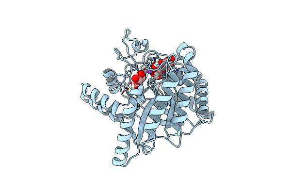
Magnetic Effects Of Thaumatin Crystals; Observation Of The Crystal Growth Of Magneto-Archimedes Levitation And Magnetic Orientation
Organism: Thaumatococcus daniellii
Method: X-RAY DIFFRACTION Resolution:0.99 Å Release Date: 2025-02-26 Classification: PLANT PROTEIN Ligands: TLA |
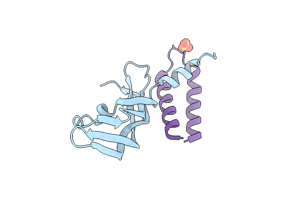 |
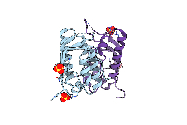
Organism: Homo sapiens, Synthetic construct
Method: X-RAY DIFFRACTION Resolution:1.45 Å Release Date: 2024-12-25 Classification: IMMUNE SYSTEM/DE NOVO PROTEIN Ligands: SO4 |
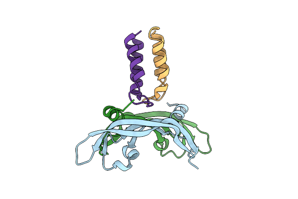 |
Organism: Homo sapiens, Synthetic construct
Method: X-RAY DIFFRACTION Resolution:1.46 Å Release Date: 2024-12-11 Classification: IMMUNE SYSTEM/DE NOVO PROTEIN |
 |
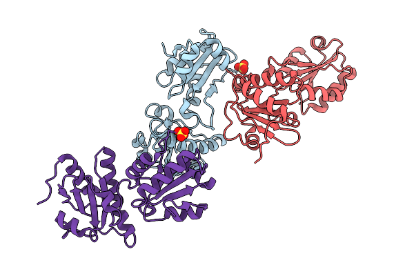
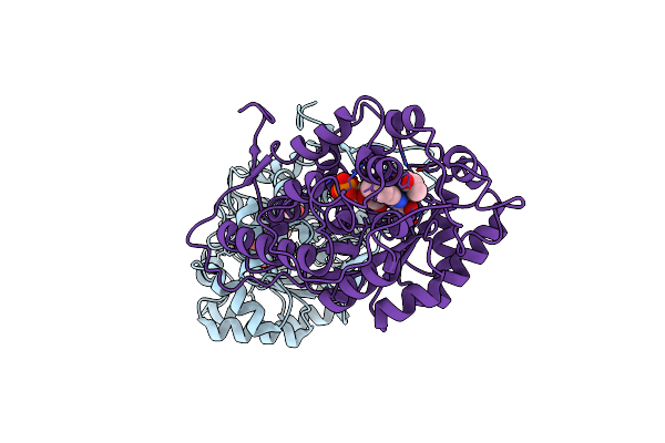
Complex Structure Of Catalytic, Small, And A Partial Electron Transfer Subunits From Burkholderia Cepacia Fad Glucose Dehydrogenase
Organism: Burkholderia cepacia
Method: X-RAY DIFFRACTION Resolution:3.00 Å Release Date: 2022-12-14 Classification: OXIDOREDUCTASE Ligands: FAD, F3S, HEM |
 |
Organism: Aerococcus viridans
Method: X-RAY DIFFRACTION Resolution:1.33 Å Release Date: 2022-06-15 Classification: OXIDOREDUCTASE Ligands: ACT, FMN, EDO |
 |
Organism: Saccharomyces cerevisiae s288c
Method: X-RAY DIFFRACTION Resolution:2.02 Å Release Date: 2022-05-18 Classification: LIPID BINDING PROTEIN Ligands: SO4 |
 |
Organism: Aerococcus viridans
Method: X-RAY DIFFRACTION Resolution:1.30 Å Release Date: 2022-03-23 Classification: OXIDOREDUCTASE Ligands: FMN, 2OP |
 |
Organism: Aerococcus viridans
Method: X-RAY DIFFRACTION Resolution:1.38 Å Release Date: 2022-03-23 Classification: OXIDOREDUCTASE Ligands: FMN, LAC |
 |
Organism: Aerococcus viridans
Method: X-RAY DIFFRACTION Resolution:1.41 Å Release Date: 2022-03-23 Classification: OXIDOREDUCTASE Ligands: FMN, PYR |
 |
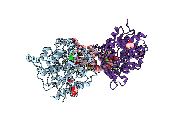
Crystal Structure Of Dipeptidyl Peptidase 11 (Dpp11) With Citrate From Porphyromonas Gingivalis (Space)
Organism: Porphyromonas gingivalis (strain atcc 33277 / dsm 20709 / cip 103683 / jcm 12257 / nctc 11834 / 2561)
Method: X-RAY DIFFRACTION Resolution:1.50 Å Release Date: 2019-10-02 Classification: HYDROLASE Ligands: PEG, CIT, GOL, K |
 |
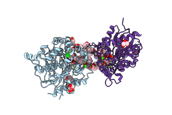
Crystal Structure Of Dipeptidyl Peptidase 11 (Dpp11) With Sh-5 From Porphyromonas Gingivalis (Space)
Organism: Porphyromonas gingivalis (strain atcc 33277 / dsm 20709 / cip 103683 / jcm 12257 / nctc 11834 / 2561)
Method: X-RAY DIFFRACTION Resolution:2.39 Å Release Date: 2019-10-02 Classification: HYDROLASE Ligands: GOL, C8O |