
Search Count: 6
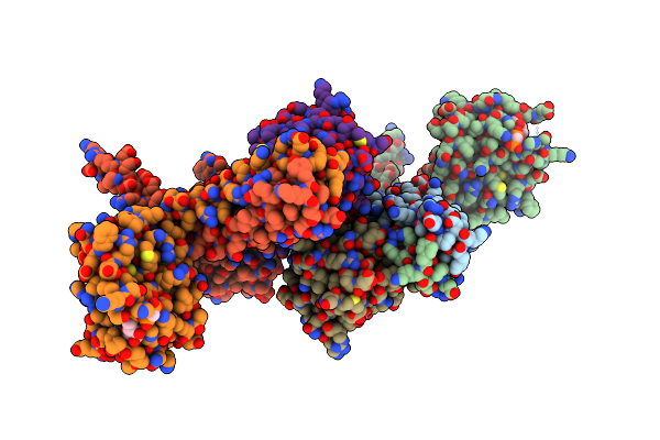 |
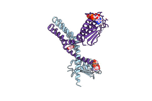
Crystal Structure Of The Desk:Desr-Q10A Complex In The Phosphotransfer State
Organism: Bacillus subtilis
Method: X-RAY DIFFRACTION Resolution:3.41 Å Release Date: 2022-11-16 Classification: TRANSFERASE Ligands: ACP, MG |
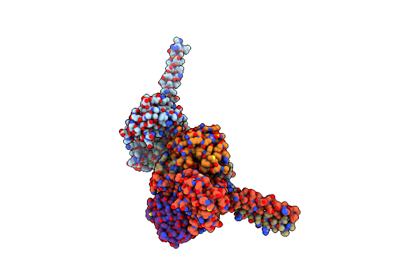 |
Organism: Bacillus subtilis
Method: X-RAY DIFFRACTION Resolution:2.52 Å Release Date: 2022-11-16 Classification: TRANSFERASE Ligands: ACP, MG, BEF, SO4 |
 |
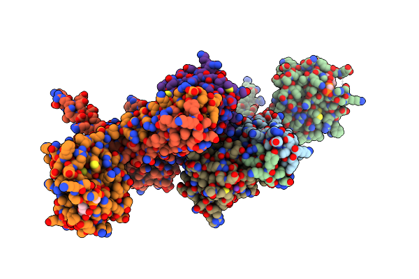
Crystal Structure Of The Desk-Desr Complex In The Phosphotransfer State With Low Mg2+ (20 Mm)
Organism: Bacillus subtilis, Bacillus subtilis (strain 168)
Method: X-RAY DIFFRACTION Resolution:3.20 Å Release Date: 2016-12-21 Classification: TRANSFERASE Ligands: ACP, MG, K |
 |
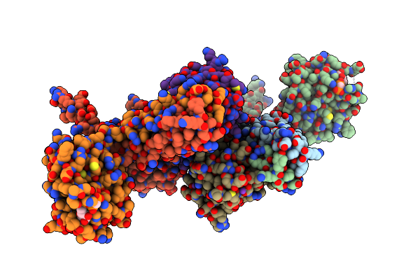
Crystal Structure Of The Desk-Desr Complex In The Phosphotransfer State With High Mg2+ (150 Mm)
Organism: Bacillus subtilis, Bacillus subtilis (strain 168)
Method: X-RAY DIFFRACTION Resolution:2.90 Å Release Date: 2016-12-21 Classification: TRANSFERASE/GENE REGULATION Ligands: ACP, MG, K |
 |
Crystal Structure Of The Desk-Desr Complex In The Phosphotransfer State With High Mg2+ (150 Mm) And Bef3
Organism: Bacillus subtilis, Bacillus subtilis (strain 168)
Method: X-RAY DIFFRACTION Resolution:3.15 Å Release Date: 2016-12-21 Classification: TRANSFERASE Ligands: ACP, MG, K |
 |
Organism: Bacillus subtilis
Method: X-RAY DIFFRACTION Resolution:3.16 Å Release Date: 2016-12-21 Classification: TRANSFERASE Ligands: ACP, MG, GOL |