
Search Count: 670
 |
Organism: Camelus dromedarius
Method: X-RAY DIFFRACTION Resolution:2.86 Å Release Date: 2026-01-28 Classification: IMMUNE SYSTEM Ligands: PG4 |
 |
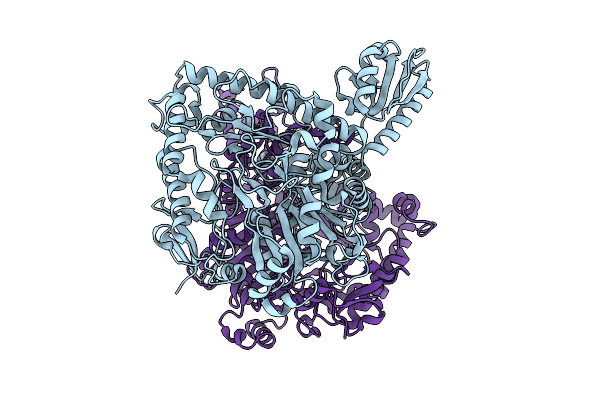
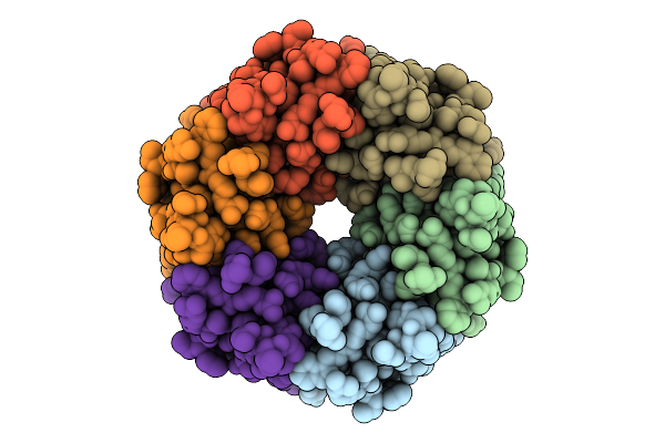
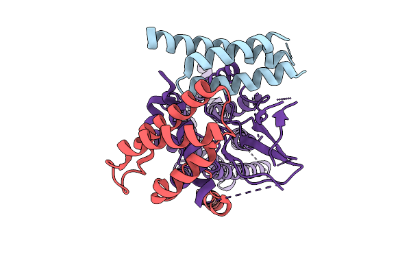
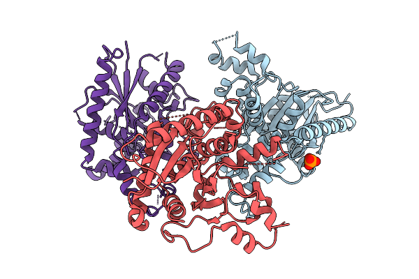
Organism: Lacticaseibacillus rhamnosus
Method: X-RAY DIFFRACTION Resolution:2.09 Å Release Date: 2025-12-24 Classification: LYASE |
 |
Organism: Lacticaseibacillus rhamnosus
Method: X-RAY DIFFRACTION Resolution:2.64 Å Release Date: 2025-12-24 Classification: LYASE |
 |
Organism: Lacticaseibacillus rhamnosus
Method: X-RAY DIFFRACTION Resolution:2.69 Å Release Date: 2025-12-24 Classification: LYASE Ligands: PLP |
 |
Organism: Lacticaseibacillus rhamnosus
Method: X-RAY DIFFRACTION Resolution:1.74 Å Release Date: 2025-12-24 Classification: LYASE |
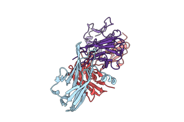 |
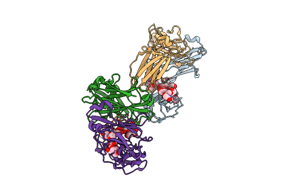
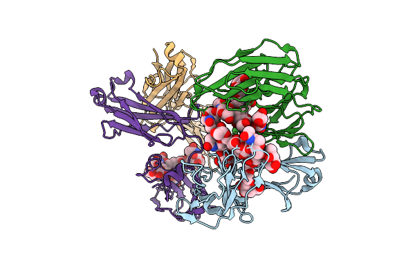
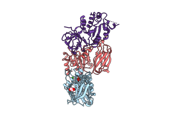
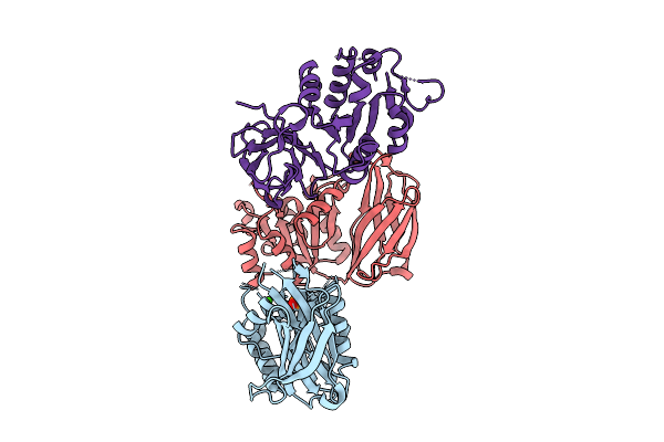
Crystal Structure Of Pilra In Complex With Fab Portion Of Antagonist Antibody
Organism: Homo sapiens
Method: X-RAY DIFFRACTION Resolution:2.58 Å Release Date: 2025-12-17 Classification: SIGNALING PROTEIN |
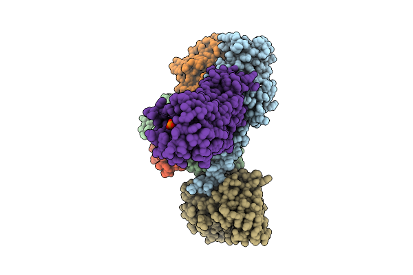 |
Organism: Homo sapiens
Method: ELECTRON MICROSCOPY Resolution:3.73 Å Release Date: 2025-11-26 Classification: MEMBRANE PROTEIN Ligands: A1L69 |
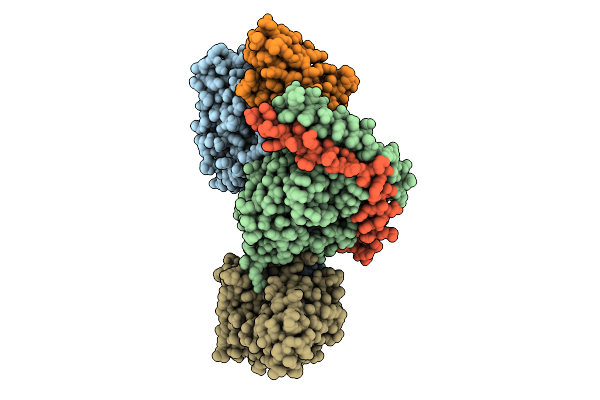 |
Organism: Homo sapiens
Method: ELECTRON MICROSCOPY Resolution:3.25 Å Release Date: 2025-11-26 Classification: MEMBRANE PROTEIN Ligands: S1P |
 |
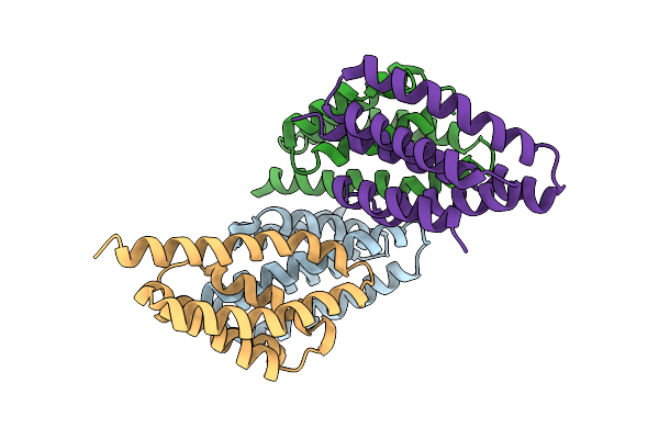
Organism: Bacillus subtilis subsp. subtilis str. 168
Method: ELECTRON MICROSCOPY Resolution:3.05 Å Release Date: 2025-11-12 Classification: STRUCTURAL PROTEIN |
 |
Organism: Aequorea
Method: X-RAY DIFFRACTION Resolution:2.37 Å Release Date: 2025-11-12 Classification: FLUORESCENT PROTEIN Ligands: CIT, 1PE, SO4 |
 |
Organism: Homo sapiens
Method: X-RAY DIFFRACTION Resolution:4.58 Å Release Date: 2025-10-29 Classification: PROTEIN BINDING |
 |
Organism: Synthetic construct
Method: X-RAY DIFFRACTION Resolution:2.07 Å Release Date: 2025-10-29 Classification: DE NOVO PROTEIN Ligands: ZN |
 |
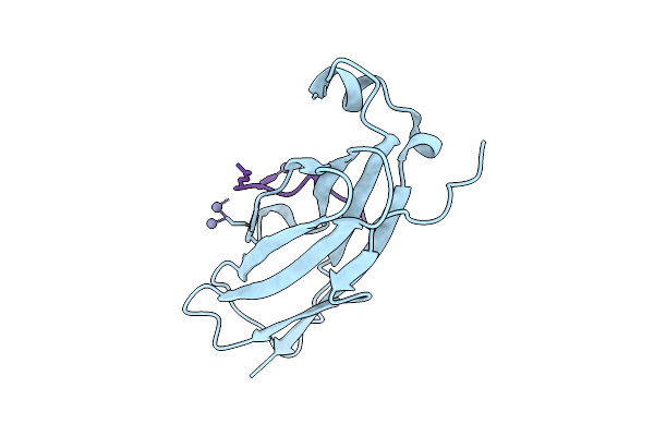
Organism: Homo sapiens
Method: X-RAY DIFFRACTION Resolution:3.12 Å Release Date: 2025-10-22 Classification: IMMUNE SYSTEM Ligands: GAL |
 |
Organism: Homo sapiens
Method: X-RAY DIFFRACTION Resolution:3.00 Å Release Date: 2025-10-22 Classification: IMMUNE SYSTEM Ligands: FUL |
 |
Organism: Homo sapiens, Synthetic construct
Method: ELECTRON MICROSCOPY Release Date: 2025-10-22 Classification: MEMBRANE PROTEIN |
 |
Organism: Synthetic construct
Method: X-RAY DIFFRACTION Resolution:3.49 Å Release Date: 2025-10-22 Classification: DE NOVO PROTEIN |
 |
Crystal Structure Of Nir2 C-Terminal Domain In Complex With Phosphatidic Acid
Organism: Mus musculus
Method: X-RAY DIFFRACTION Resolution:2.15 Å Release Date: 2025-10-15 Classification: LIPID BINDING PROTEIN Ligands: 44E, DW3, CA |
 |
Organism: Mus musculus
Method: X-RAY DIFFRACTION Resolution:2.05 Å Release Date: 2025-10-15 Classification: LIPID BINDING PROTEIN Ligands: ZN |
 |
Organism: Homo sapiens
Method: X-RAY DIFFRACTION Resolution:2.80 Å Release Date: 2025-10-15 Classification: LIPID BINDING PROTEIN Ligands: PO4 |
 |
Organism: Mus musculus
Method: X-RAY DIFFRACTION Resolution:2.40 Å Release Date: 2025-10-15 Classification: LIPID BINDING PROTEIN Ligands: CA, PO4 |