
Search Count: 23
 |
Organism: Danio rerio
Method: ELECTRON MICROSCOPY Release Date: 2023-03-22 Classification: MEMBRANE PROTEIN Ligands: NAG, PIO, PX4, SY9 |
 |
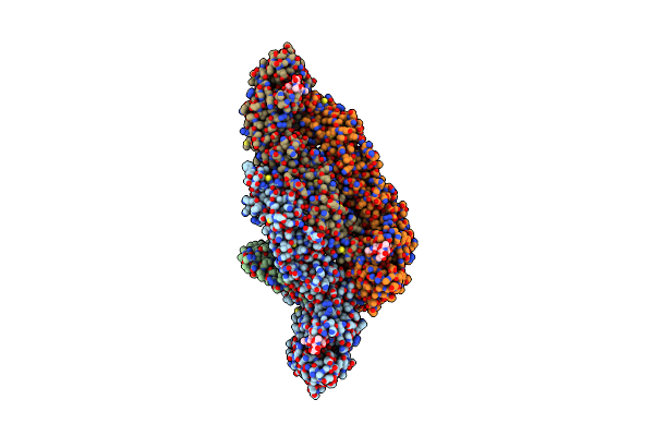
Organism: Danio rerio
Method: ELECTRON MICROSCOPY Release Date: 2023-03-22 Classification: MEMBRANE PROTEIN Ligands: GLY, NAG |
 |
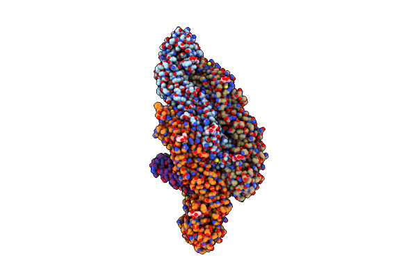
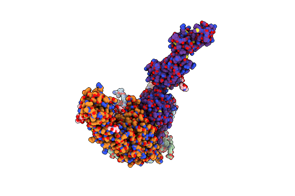
Alpha1/Betab Heteromeric Glycine Receptor In 1 Mm Glycine 20 Um Ivermectin State
Organism: Danio rerio
Method: ELECTRON MICROSCOPY Release Date: 2023-03-22 Classification: MEMBRANE PROTEIN Ligands: GLY, NAG, IVM, PX4, PLM, D10 |
 |
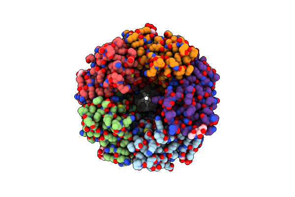
Organism: Spondweni virus
Method: ELECTRON MICROSCOPY Release Date: 2021-01-20 Classification: VIRUS Ligands: NAG |
 |
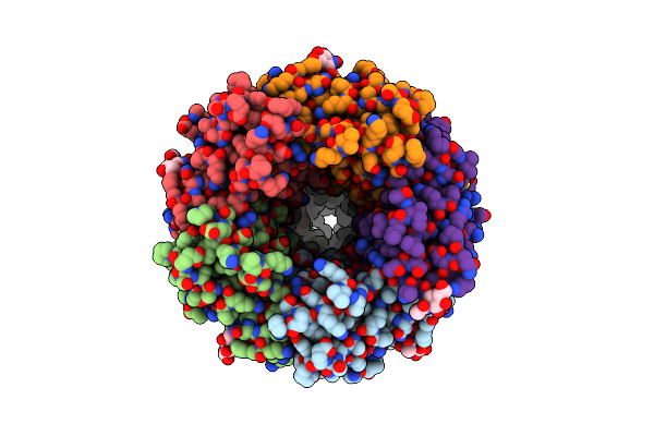
Organism: Spondweni virus
Method: ELECTRON MICROSCOPY Release Date: 2021-01-20 Classification: VIRUS Ligands: NAG |
 |
Organism: Dengue virus 2
Method: ELECTRON MICROSCOPY Release Date: 2021-01-20 Classification: VIRUS Ligands: NAG |
 |
Organism: Spondweni virus
Method: ELECTRON MICROSCOPY Release Date: 2021-01-20 Classification: VIRUS Ligands: 3PE |
 |
Organism: Spondweni virus
Method: ELECTRON MICROSCOPY Release Date: 2021-01-20 Classification: VIRUS Ligands: NAG |
 |
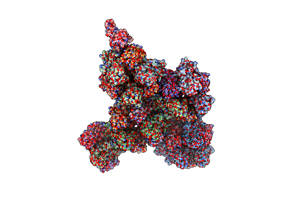
Atomic Models Of P1, P4 C-Terminal Fragment And P8 Fitted In The Bacteriophage Phi6 Nucleocapsid Reconstructed With Icosahedral Symmetry
Organism: Pseudomonas phage phi6
Method: ELECTRON MICROSCOPY Release Date: 2019-06-12 Classification: VIRUS |
 |
Organism: Haloarcula hispanica virus sh1
Method: ELECTRON MICROSCOPY Release Date: 2019-04-10 Classification: VIRUS |
 |
Organism: Foot-and-mouth disease virus
Method: ELECTRON MICROSCOPY Release Date: 2017-06-21 Classification: VIRUS |
 |
Organism: Foot-and-mouth disease virus
Method: ELECTRON MICROSCOPY Release Date: 2017-06-21 Classification: VIRUS |
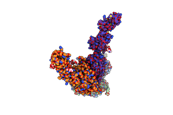 |
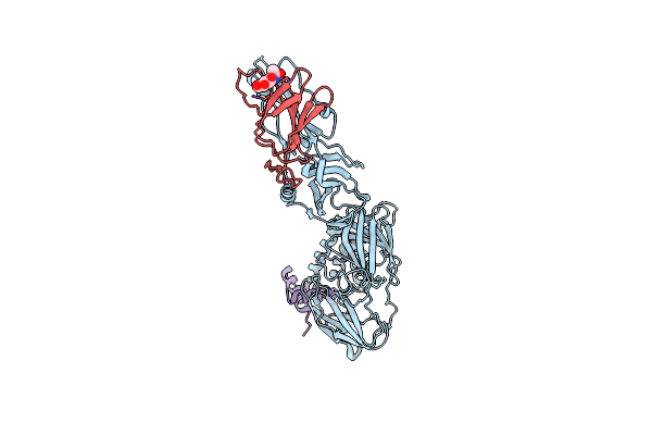
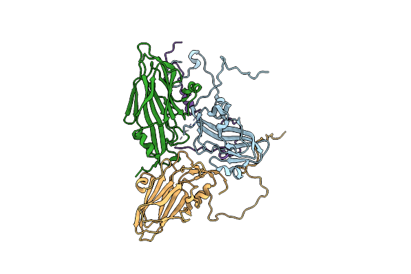
Localised Reconstruction Of Alpha V Beta 6 Bound To Foot And Mouth Disease Virus O Panasia - Pose A.
Organism: Foot-and-mouth disease virus - type o, Homo sapiens
Method: ELECTRON MICROSCOPY Release Date: 2017-06-21 Classification: VIRUS Ligands: MAN, NAG, CA |
 |
Localised Reconstruction Of Alpha V Beta 6 Bound To Foot And Mouth Disease Virus O Panasia - Pose A Prime.
Organism: Foot-and-mouth disease virus, Homo sapiens
Method: ELECTRON MICROSCOPY Release Date: 2017-06-21 Classification: VIRUS Ligands: MAN, NAG, CA |
 |
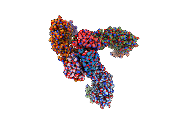
Localised Reconstruction Of Integrin Alpha V Beta 6 Bound To Foot And Mouth Disease Virus O1 Manisa - Pose A.
Organism: Foot-and-mouth disease virus, Homo sapiens
Method: ELECTRON MICROSCOPY Release Date: 2017-06-21 Classification: VIRUS Ligands: MAN, NAG, CA |
 |
Localised Reconstruction Of Integrin Alpha V Beta 6 Bound To Foot And Mouth Disease Virus O1 Manisa - Pose B.
Organism: Foot-and-mouth disease virus, Homo sapiens
Method: ELECTRON MICROSCOPY Release Date: 2017-06-21 Classification: VIRUS Ligands: MG, CA |
 |
Organism: Foot-and-mouth disease virus, Foot-and-mouth disease virus - type o
Method: X-RAY DIFFRACTION Resolution:2.30 Å Release Date: 2017-06-14 Classification: VIRUS |
 |
Organism: Pseudomonas phage phi6
Method: ELECTRON MICROSCOPY Release Date: 2017-03-22 Classification: VIRUS |
 |
Atomic Structure Fitted Into A Localized Reconstruction Of Bacteriophage Phi6 Packaging Hexamer P4
Organism: Pseudomonas phage phi6
Method: ELECTRON MICROSCOPY Release Date: 2017-03-22 Classification: HYDROLASE Ligands: CA, ADP |
 |
Atomic Structure Of P4 Packaging Enzyme Fitted Into A Localized Reconstruction Of Bacteriophage Phi6 Vertex
Organism: Pseudomonas phage phi6
Method: ELECTRON MICROSCOPY Resolution:9.10 Å Release Date: 2017-03-22 Classification: HYDROLASE Ligands: CA, ADP |