Search Count: 28
 |
Organism: Paenibacillus sp. 598k
Method: X-RAY DIFFRACTION Resolution:2.00 Å Release Date: 2017-07-26 Classification: HYDROLASE, TRANSFERASE Ligands: CA, NI, MG, SO4, MES, EDO |
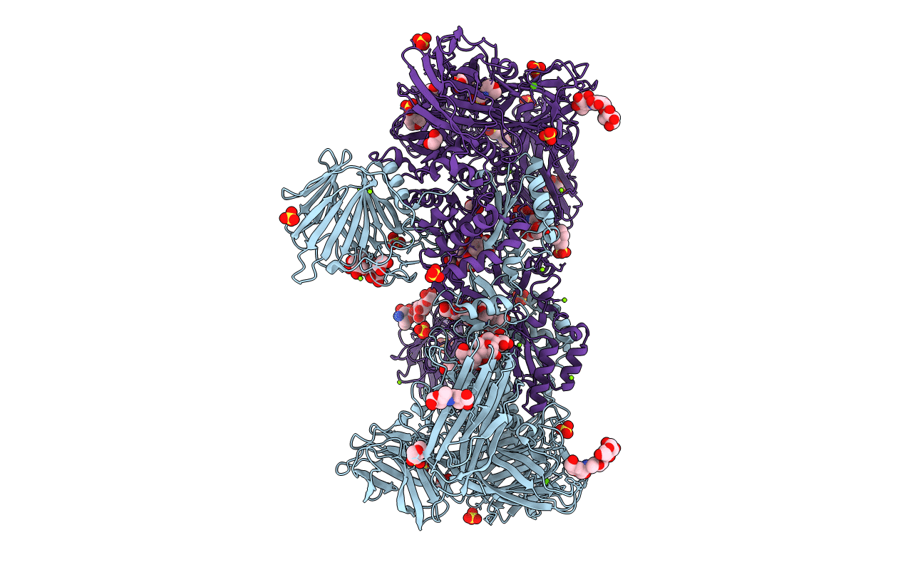 |
Crystal Structure Of Paenibacillus Sp. 598K Alpha-1,6-Glucosyltransferase Complexed With Acarbose
Organism: Paenibacillus sp. 598k
Method: X-RAY DIFFRACTION Resolution:2.40 Å Release Date: 2017-07-26 Classification: HYDROLASE, TRANSFERASE Ligands: CA, NI, MG, SO4, MES, EDO |
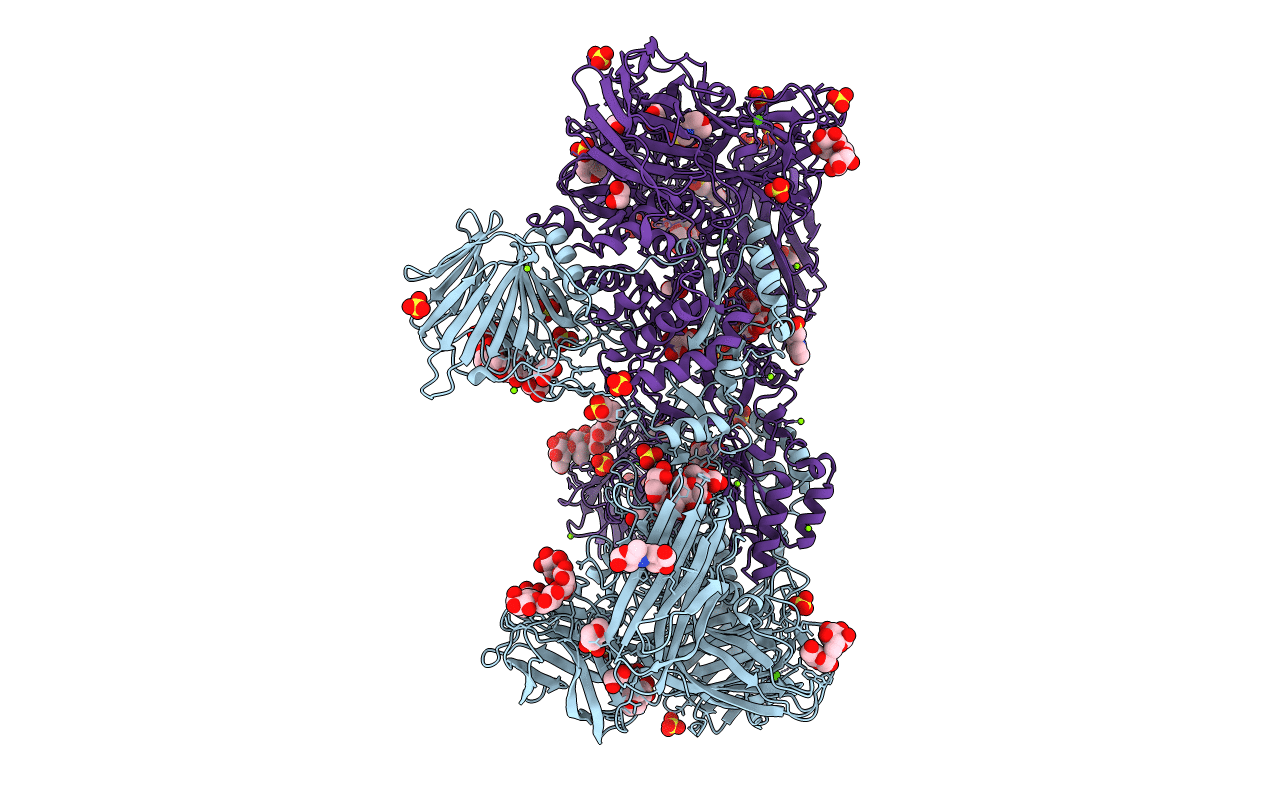 |
Crystal Structure Of Paenibacillus Sp. 598K Alpha-1,6-Glucosyltransferase Complexed With Maltohexaose
Organism: Paenibacillus sp. 598k
Method: X-RAY DIFFRACTION Resolution:1.95 Å Release Date: 2017-07-26 Classification: HYDROLASE, TRANSFERASE Ligands: CA, BGC, GLC, NI, MG, SO4, MES, EDO |
 |
Crystal Structure Of Paenibacillus Sp. 598K Alpha-1,6-Glucosyltransferase Complexed With Isomaltohexaose
Organism: Paenibacillus sp. 598k
Method: X-RAY DIFFRACTION Resolution:1.95 Å Release Date: 2017-07-26 Classification: HYDROLASE, TRANSFERASE Ligands: CA, BGC, GLC, NI, MG, SO4, MES, EDO |
 |
Crystal Structure Of Paenibacillus Sp. 598K Alpha-1,6-Glucosyltransferase, Terbium Derivative
Organism: Paenibacillus sp. 598k
Method: X-RAY DIFFRACTION Resolution:2.40 Å Release Date: 2017-07-26 Classification: HYDROLASE, TRANSFERASE Ligands: CA, NI, MG, TB, SO4, MES, EDO |
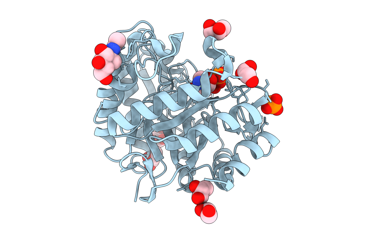 |
Crtstal Structure Of Glycoside Hydrolase Family 5 Beta-Mannanase From Talaromyces Trachyspermus
Organism: Talaromyces trachyspermus
Method: X-RAY DIFFRACTION Resolution:1.60 Å Release Date: 2014-07-23 Classification: HYDROLASE Ligands: NAG, TRS, GOL, PO4 |
 |
Organism: Streptomyces coelicolor
Method: X-RAY DIFFRACTION Resolution:1.40 Å Release Date: 2014-02-05 Classification: HYDROLASE Ligands: CA, CL, TRS |
 |
Crystal Structure Of Streptomyces Coelicolor Alpha-L-Arabinofuranosidase Ethylmercury Derivative
Organism: Streptomyces coelicolor
Method: X-RAY DIFFRACTION Resolution:1.90 Å Release Date: 2014-02-05 Classification: HYDROLASE Ligands: CA, TRS, EMC |
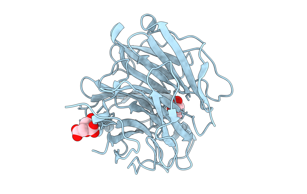 |
Crystal Structure Of Streptomyces Coelicolor Alpha-L-Arabinofuranosidase In Complex With L-Arabinose
Organism: Streptomyces coelicolor
Method: X-RAY DIFFRACTION Resolution:1.90 Å Release Date: 2014-02-05 Classification: HYDROLASE Ligands: CA, CL, FUB, CIT |
 |
Crystal Structure Of Streptomyces Coelicolor Alpha-L-Arabinofuranosidase In Complex With Xylotriose
Organism: Streptomyces coelicolor
Method: X-RAY DIFFRACTION Resolution:2.00 Å Release Date: 2014-02-05 Classification: HYDROLASE Ligands: CA, CL, TRS, CIT |
 |
Crystal Structure Of Streptomyces Coelicolor Alpha-L-Arabinofuranosidase In Complex With Xylohexaose
Organism: Streptomyces coelicolor
Method: X-RAY DIFFRACTION Resolution:2.10 Å Release Date: 2014-02-05 Classification: HYDROLASE Ligands: CA, CL, TRS, CIT |
 |
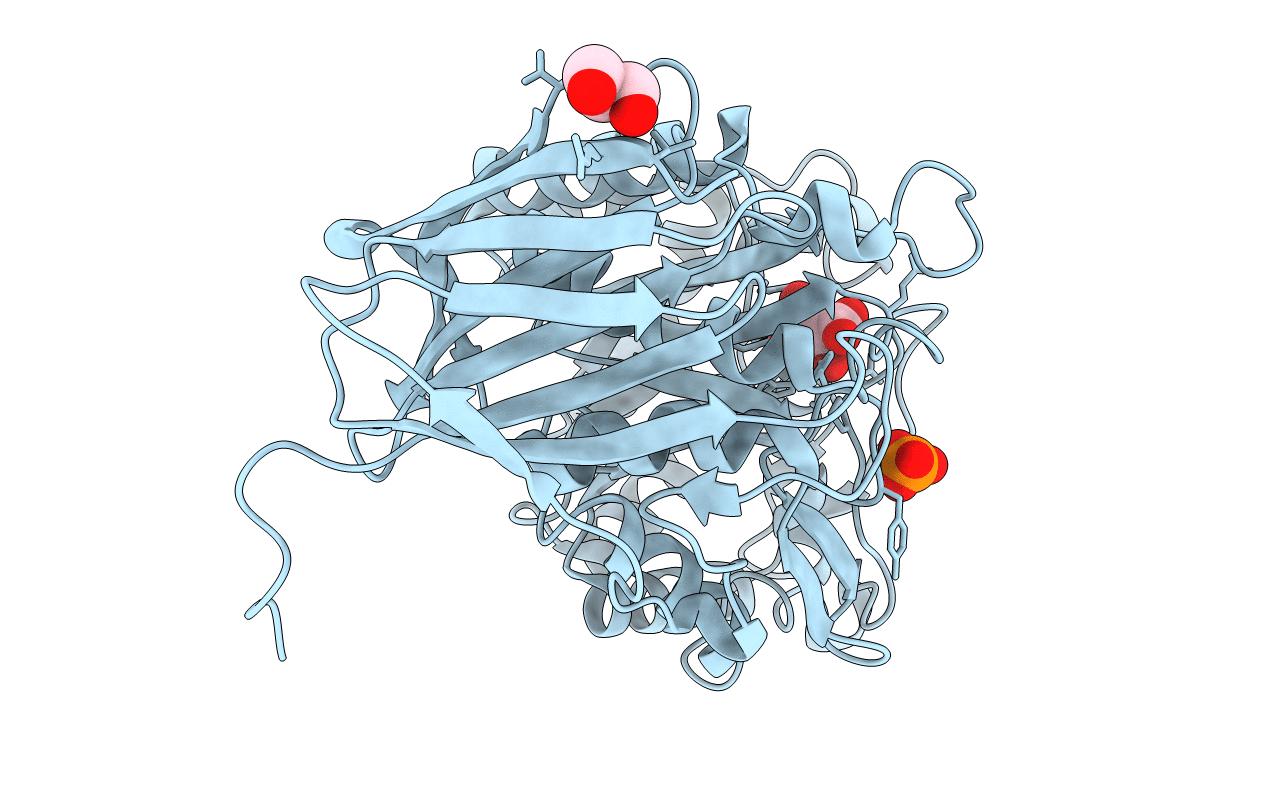
Organism: Acidobacterium capsulatum
Method: X-RAY DIFFRACTION Resolution:1.50 Å Release Date: 2012-02-22 Classification: HYDROLASE Ligands: PO4, GOL |
 |
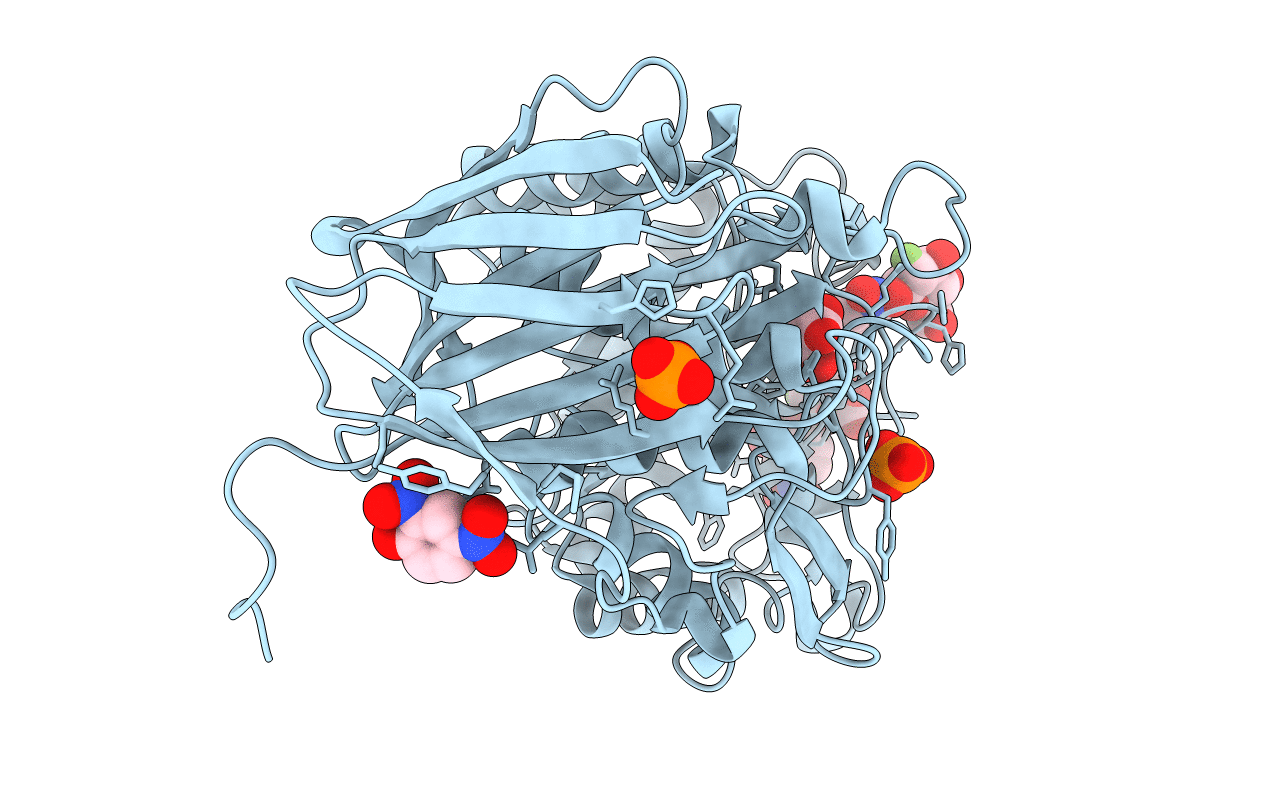
Crystal Structure Of Beta-Glucuronidase From Acidobacterium Capsulatum In Complex With D-Glucuronic Acid
Organism: Acidobacterium capsulatum
Method: X-RAY DIFFRACTION Resolution:1.80 Å Release Date: 2012-02-22 Classification: HYDROLASE Ligands: BDP, PO4, GOL |
 |
Crystal Structure Of Beta-Glucuronidase From Acidobacterium Capsulatum Covalent-Bonded With 2-Deoxy-2-Fluoro-D-Glucuronic Acid
Organism: Acidobacterium capsulatum
Method: X-RAY DIFFRACTION Resolution:1.90 Å Release Date: 2012-02-22 Classification: HYDROLASE Ligands: GUZ, GUF, DNF, PO4 |
 |
The Structure And Function Of An Arabinan-Specific Alpha-1,2-Arabinofuranosidase Identified From Screening The Activities Of Bacterial Gh43 Glycoside Hydrolases
Organism: Cellvibrio japonicus
Method: X-RAY DIFFRACTION Resolution:2.99 Å Release Date: 2011-02-16 Classification: HYDROLASE Ligands: SO4, CA, TAM |
 |
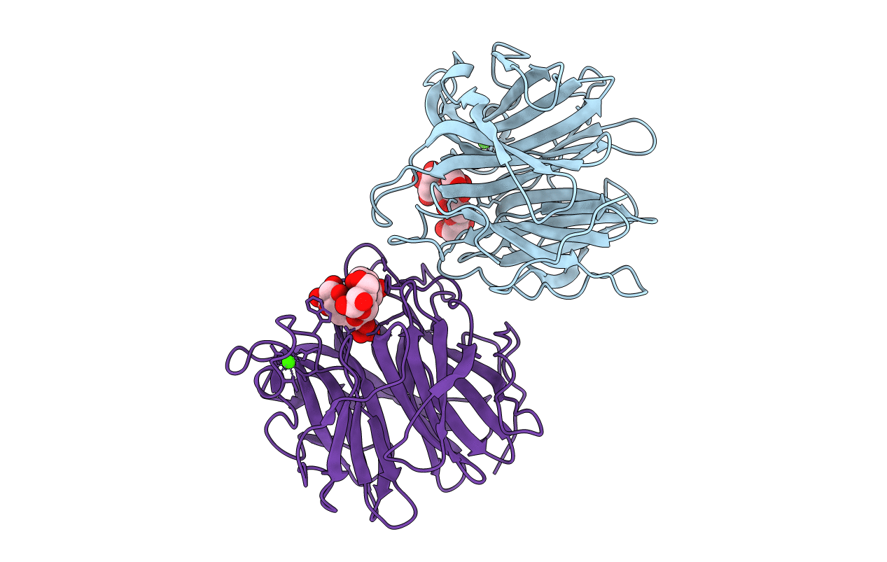
The Structure And Function Of An Arabinan-Specific Alpha-1,2-Arabinofuranosidase Identified From Screening The Activities Of Bacterial Gh43 Glycoside Hydrolases
Organism: Cellvibrio japonicus
Method: X-RAY DIFFRACTION Resolution:1.64 Å Release Date: 2011-02-16 Classification: HYDROLASE Ligands: CA, ACT |
 |
The Structure And Function Of An Arabinan-Specific Alpha-1,2-Arabinofuranosidase Identified From Screening The Activities Of Bacterial Gh43 Glycoside Hydrolases
Organism: Cellvibrio japonicus
Method: X-RAY DIFFRACTION Resolution:1.79 Å Release Date: 2011-02-16 Classification: HYDROLASE Ligands: CA, EDO |
 |
Organism: Streptomyces avermitilis
Method: X-RAY DIFFRACTION Resolution:2.20 Å Release Date: 2010-08-25 Classification: HYDROLASE Ligands: CL, NA, GOL |
 |
Crystal Structure Of Exo-1,5-Alpha-L-Arabinofuranosidase Complexed With Alpha-1,5-L-Arabinofuranobiose
Organism: Streptomyces avermitilis
Method: X-RAY DIFFRACTION Resolution:1.80 Å Release Date: 2010-08-25 Classification: HYDROLASE Ligands: CL, NA, AHR, GOL |
 |
Crystal Structure Of Exo-1,5-Alpha-L-Arabinofuranosidase Complexed With Alpha-1,5-L-Arabinofuranotriose
Organism: Streptomyces avermitilis
Method: X-RAY DIFFRACTION Resolution:1.70 Å Release Date: 2010-08-25 Classification: HYDROLASE Ligands: CL, NA, AHR, GOL |

