
Search Count: 25
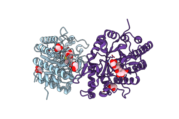 |
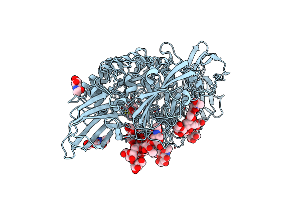
Crystal Structure Of Xyn11 Double Mutant L271S, K275H From Psedothermotoga Thermarum
Organism: Pseudothermotoga thermarum dsm 5069
Method: X-RAY DIFFRACTION Resolution:2.10 Å Release Date: 2023-10-25 Classification: HYDROLASE Ligands: GOL, IPT |
 |
Organism: Pseudothermotoga thermarum dsm 5069
Method: X-RAY DIFFRACTION Resolution:1.80 Å Release Date: 2021-06-09 Classification: HYDROLASE Ligands: GOL, IPT |
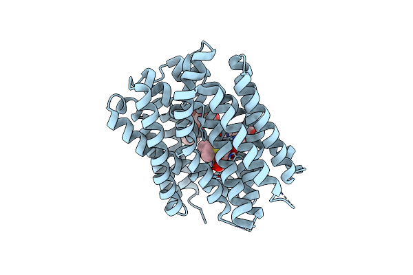 |
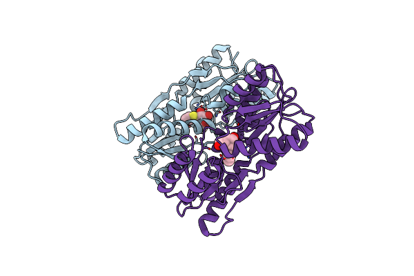
Crystal Structure Of Drug:Proton Antiporter-1 (Dha1) Family Sotb, In The Inward-Occluded Conformation
Organism: Escherichia coli (strain k12)
Method: X-RAY DIFFRACTION Resolution:3.06 Å Release Date: 2020-07-29 Classification: PROTEIN TRANSPORT Ligands: BNG, IPT |
 |
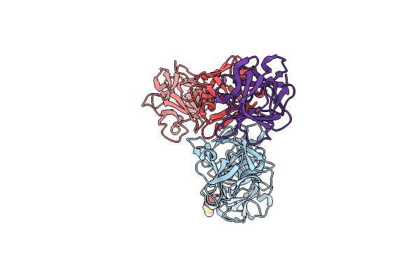
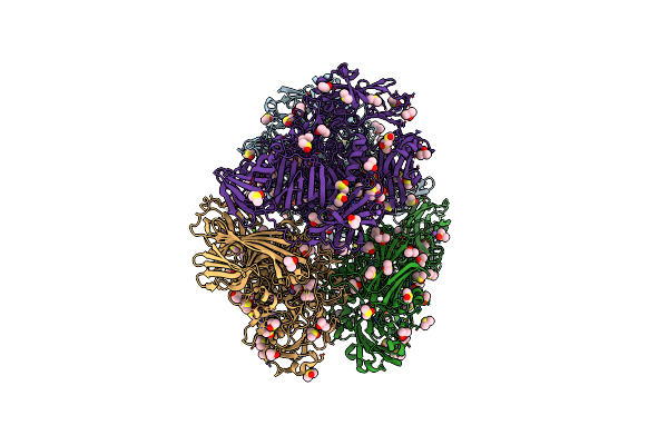
Cold-Adapted Beta-D-Galactosidase From Arthrobacter Sp. 32Cb In Complex With Iptg
Organism: Arthrobacter sp. 32cb
Method: X-RAY DIFFRACTION Resolution:2.27 Å Release Date: 2019-09-11 Classification: HYDROLASE Ligands: IPT, ACT, NA, FMT |
 |
Organism: Clostridium botulinum
Method: X-RAY DIFFRACTION Resolution:2.18 Å Release Date: 2015-09-02 Classification: PROTEIN BINDING Ligands: IPT |
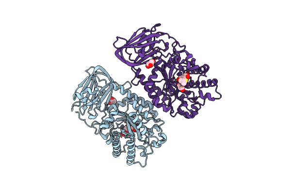 |
Crystal Structure Of An A-L-Fucosidase Gh29 From Bacteroides Thetaiotaomicron (Bt2192) In Complex With Iptg
Organism: Bacteroides thetaiotaomicron
Method: X-RAY DIFFRACTION Resolution:2.35 Å Release Date: 2014-02-26 Classification: HYDROLASE Ligands: GOL, IPT |
 |
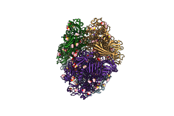
Crystal Structure Of Ct1,3Gal43A In Complex With Isopropy-Beta-D-Thiogalactoside
Organism: Clostridium thermocellum
Method: X-RAY DIFFRACTION Resolution:3.00 Å Release Date: 2012-12-05 Classification: SUGAR BINDING PROTEIN Ligands: IPT, GOL |
 |
Organism: Escherichia coli
Method: X-RAY DIFFRACTION Resolution:2.00 Å Release Date: 2012-04-11 Classification: HYDROLASE Ligands: MG, NA, DMS, IPT |
 |
Organism: Escherichia coli
Method: X-RAY DIFFRACTION Resolution:2.05 Å Release Date: 2012-04-11 Classification: HYDROLASE Ligands: MG, NA, IPT, DMS |
 |
Organism: Escherichia coli
Method: X-RAY DIFFRACTION Resolution:2.55 Å Release Date: 2012-04-11 Classification: HYDROLASE Ligands: IPT, MG, NA, DMS |
 |
Organism: Escherichia coli
Method: X-RAY DIFFRACTION Resolution:2.00 Å Release Date: 2012-01-18 Classification: HYDROLASE Ligands: MG, NA, IPT, DMS |
 |
Organism: Escherichia coli
Method: X-RAY DIFFRACTION Resolution:2.40 Å Release Date: 2012-01-18 Classification: HYDROLASE Ligands: MG, NA, IPT, DMS |
 |
Organism: Escherichia coli
Method: X-RAY DIFFRACTION Resolution:2.60 Å Release Date: 2012-01-18 Classification: HYDROLASE Ligands: IPT, MG, NA, DMS |
 |
Organism: Escherichia coli
Method: X-RAY DIFFRACTION Resolution:1.90 Å Release Date: 2011-03-16 Classification: HYDROLASE Ligands: IPT, MG, NA, DMS |
 |
Organism: Trichoderma reesei
Method: X-RAY DIFFRACTION Resolution:1.75 Å Release Date: 2011-03-16 Classification: HYDROLASE Ligands: IPT, NAG |
 |
Organism: Escherichia coli
Method: X-RAY DIFFRACTION Resolution:2.20 Å Release Date: 2010-05-12 Classification: HYDROLASE Ligands: IPT, MG, NA, DMS |
 |
Organism: Escherichia coli k12
Method: X-RAY DIFFRACTION Resolution:1.80 Å Release Date: 2008-10-28 Classification: HYDROLASE Ligands: MG, NA, IPT, DMS |
 |
Organism: Escherichia coli
Method: X-RAY DIFFRACTION Resolution:2.00 Å Release Date: 2007-06-19 Classification: TRANSCRIPTION Ligands: IPT |
 |
Organism: Escherichia coli
Method: X-RAY DIFFRACTION Resolution:1.60 Å Release Date: 2004-06-15 Classification: HYDROLASE Ligands: MG, NA, IPT, DMS |
 |
Organism: Unidentified
Method: X-RAY DIFFRACTION Resolution:1.60 Å Release Date: 2003-06-03 Classification: DE NOVO PROTEIN Ligands: PB, ACT, CL, NA, MG, IPT |