
Search Count: 65
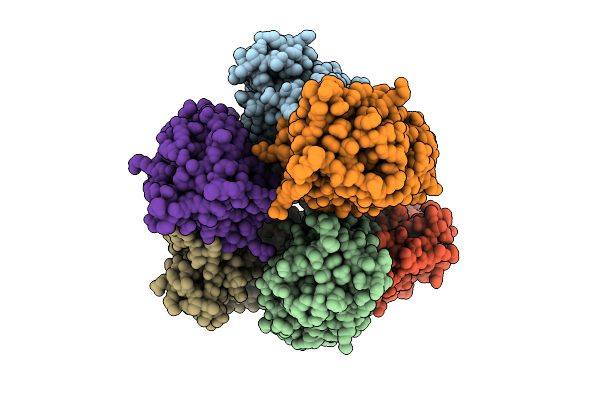 |
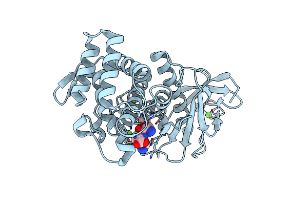
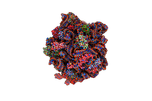
Crystal Structure Of Non-Haem Diiron Azetidine Synthase From Streptomyces Cacaoi Var. Asoensis Complexed With Iron, L-Isoleucine And Molecular Oxygen
Organism: Streptomyces cacaoi
Method: X-RAY DIFFRACTION Release Date: 2025-12-24 Classification: METAL BINDING PROTEIN Ligands: ILE, FE2, OXY |
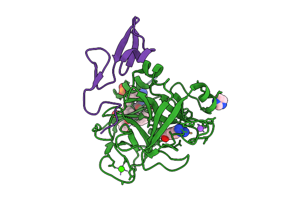 |
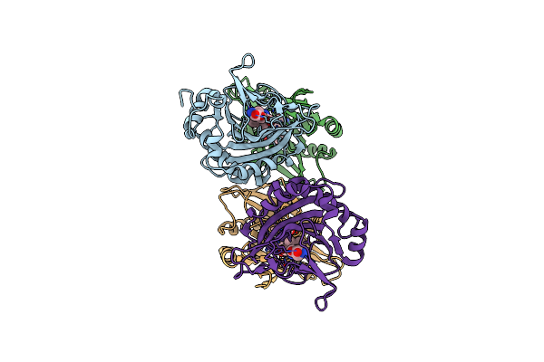
Organism: Homo sapiens, Synthetic construct
Method: X-RAY DIFFRACTION Release Date: 2025-11-26 Classification: BLOOD CLOTTING Ligands: CA, NA, CL, IMD, 0GJ, GLU, ILE, PHE, PTR |
 |
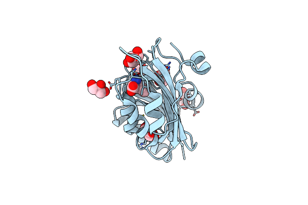
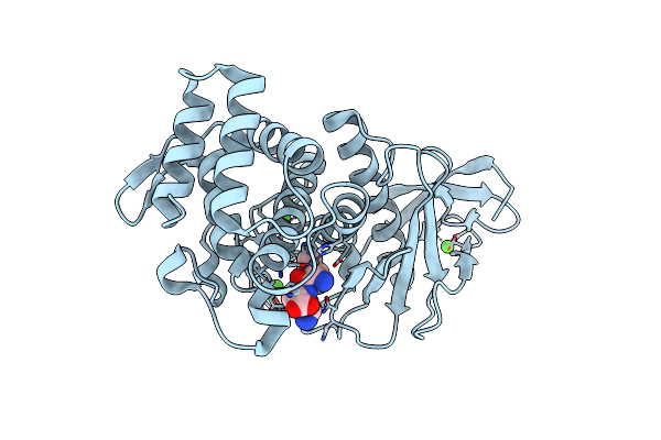
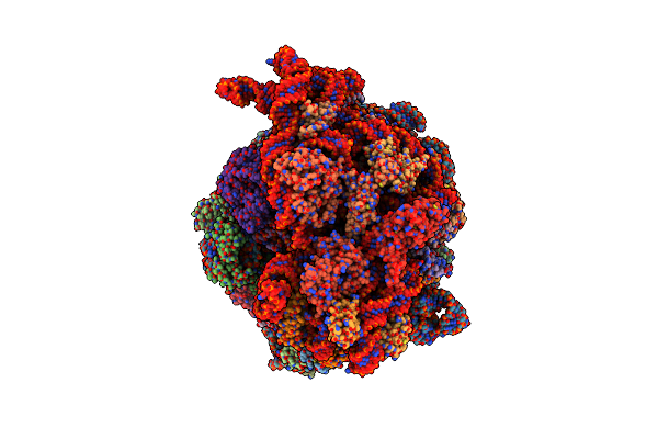
X-Ray Crystal Structure Of Streptomyces Cacaoi Polf In Complex With Iron And L-Isoleucine
Organism: Streptomyces cacaoi
Method: X-RAY DIFFRACTION Release Date: 2025-10-15 Classification: OXIDOREDUCTASE Ligands: GOL, FE2, ZN, ILE |
 |
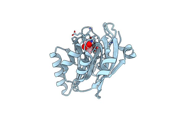
Organism: Streptomyces armeniacus
Method: X-RAY DIFFRACTION Release Date: 2025-10-08 Classification: OXIDOREDUCTASE Ligands: FE2, ILE |
 |
Organism: Streptomyces sp. z26
Method: X-RAY DIFFRACTION Release Date: 2025-10-08 Classification: OXIDOREDUCTASE Ligands: FE2, ILE |
 |
Organism: Streptomyces sp. z26
Method: X-RAY DIFFRACTION Release Date: 2025-10-08 Classification: OXIDOREDUCTASE Ligands: FE2, ILE |
 |
Organism: Streptomyces sp. nth33
Method: X-RAY DIFFRACTION Release Date: 2025-10-08 Classification: OXIDOREDUCTASE Ligands: FE2, ILE, H4B |
 |
Organism: Streptomyces sp. nth33
Method: X-RAY DIFFRACTION Release Date: 2025-10-08 Classification: OXIDOREDUCTASE Ligands: FE2, ILE |
 |
Organism: Streptomyces asoensis
Method: X-RAY DIFFRACTION Release Date: 2025-10-08 Classification: OXIDOREDUCTASE Ligands: FE2, ILE |
 |
Crystal Structure Of E. Coli Threonine Dehydratase Regulatory Domain In Complex With Isoleucine
Organism: Escherichia coli
Method: X-RAY DIFFRACTION Release Date: 2025-04-09 Classification: LYASE Ligands: ILE, GOL, ACT |
 |
Crystal Structure Of E. Coli Threonine Dehydratase Regulatory Domain F352A Mutant In Complex With Isoleucine
Organism: Escherichia coli
Method: X-RAY DIFFRACTION Release Date: 2025-04-09 Classification: LYASE Ligands: ILE, GOL |
 |
Organism: Escherichia coli o157:h7, Branchiostoma lanceolatum
Method: X-RAY DIFFRACTION Resolution:2.65 Å Release Date: 2024-12-25 Classification: FLUORESCENT PROTEIN Ligands: ILE, GOL, CL |
 |
Organism: Escherichia coli, Escherichia coli o157:h7
Method: X-RAY DIFFRACTION Resolution:1.62 Å Release Date: 2024-12-25 Classification: FLUORESCENT PROTEIN Ligands: ILE, GOL, CL |
 |
Crystal Structure Of Saccharomyces Cerevisiae Isoleucyl-Trna Synthetase In Complex With Trna(Ile) And Isoleucine
Organism: Saccharomyces cerevisiae, Escherichia coli
Method: X-RAY DIFFRACTION Resolution:2.80 Å Release Date: 2024-10-09 Classification: LIGASE/RNA Ligands: ILE, EDO, SO4 |
 |
Organism: Bacillus licheniformis
Method: X-RAY DIFFRACTION Release Date: 2024-09-04 Classification: OXIDOREDUCTASE Ligands: ILE |
 |
Organism: Bacillus thermoproteolyticus
Method: X-RAY DIFFRACTION Resolution:1.40 Å Release Date: 2024-06-05 Classification: HYDROLASE Ligands: ZN, CA, ILE, LYS |
 |
Organism: Bacillus thermoproteolyticus
Method: X-RAY DIFFRACTION Resolution:1.40 Å Release Date: 2024-06-05 Classification: HYDROLASE Ligands: ILE, LYS, ZN, CA |
 |
Organism: Bacillus thermoproteolyticus
Method: X-RAY DIFFRACTION Resolution:1.40 Å Release Date: 2024-06-05 Classification: HYDROLASE Ligands: ILE, LYS, ZN, CA |
 |
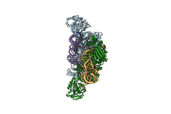
Structure Of The Escherichia Coli 70S Ribosome In Complex With Ef-Tu And Ile-Trnaile(Lau) Bound To The Cognate Aua Codon (Structure I)
Organism: Escherichia coli, Escherichia phage t4
Method: ELECTRON MICROSCOPY Release Date: 2024-03-06 Classification: RIBOSOME Ligands: MG, K, ILE, GCP, ZN |
 |
Structure Of The Escherichia Coli 70S Ribosome In Complex With Ef-Tu And Ile-Trnaile(Lau) Bound To The Near-Cognate Aug Codon (Structure Ii)
Organism: Escherichia coli, Escherichia phage t4
Method: ELECTRON MICROSCOPY Release Date: 2024-03-06 Classification: RIBOSOME Ligands: PAR, MG, ILE, GCP, ZN |