
Search Count: 15
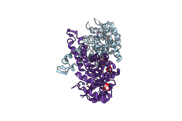 |
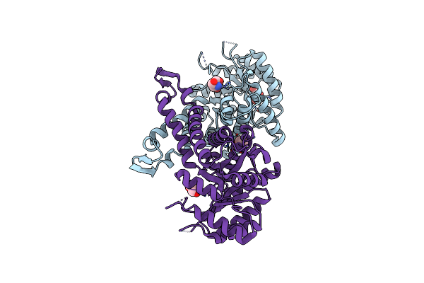
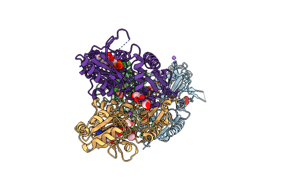
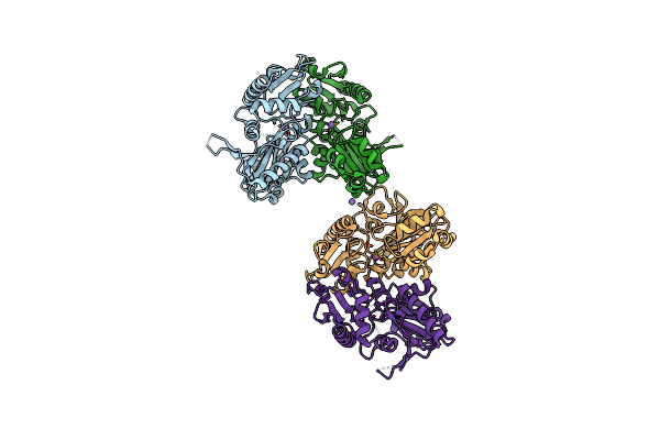
Native Structure Of Fructose 1,6-Bisphosphate Aldolase From Escherichia Coli At 1.8 Angstrom Resolution
Organism: Escherichia coli (strain k12)
Method: X-RAY DIFFRACTION Resolution:1.80 Å Release Date: 2017-07-05 Classification: LYASE Ligands: GOL, ZN, PEG |
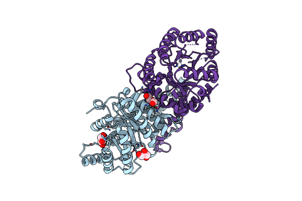 |
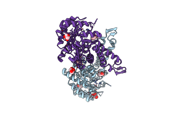
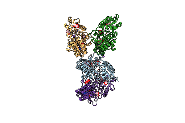
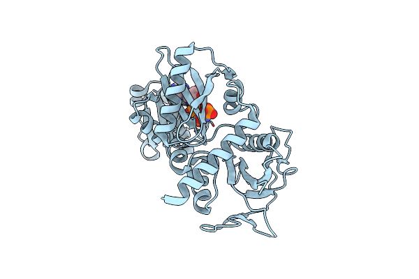
Native Structure Of Fructose 1,6-Bisphosphate Aldolase From Escherichia Coli At 2.0 Angstrom Resolution
Organism: Escherichia coli (strain k12)
Method: X-RAY DIFFRACTION Resolution:2.00 Å Release Date: 2017-07-05 Classification: LYASE Ligands: GOL, ZN, PEG |
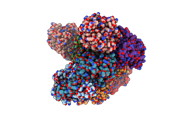 |
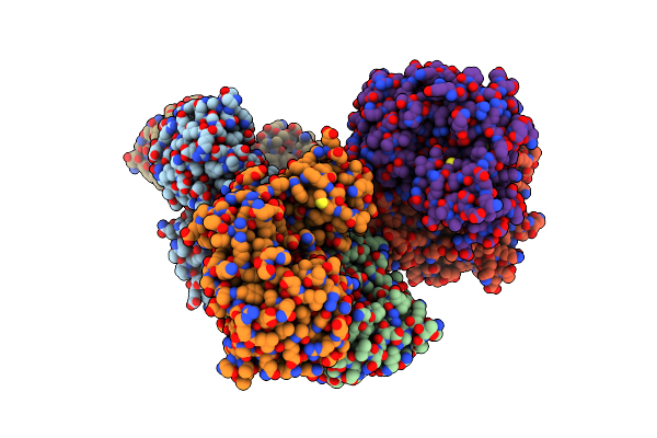
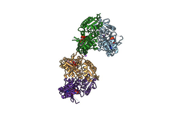
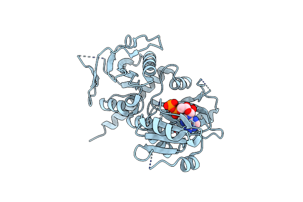
Apo Structure Of Fructose 1,6-Bisphosphate Aldolase From Escherichia Coli At 1.9 Angstrom Resolution
Organism: Escherichia coli (strain k12)
Method: X-RAY DIFFRACTION Resolution:1.90 Å Release Date: 2017-07-05 Classification: LYASE Ligands: GOL, ZN, PEG |
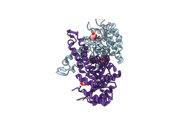 |
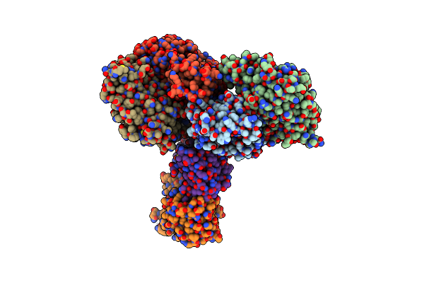
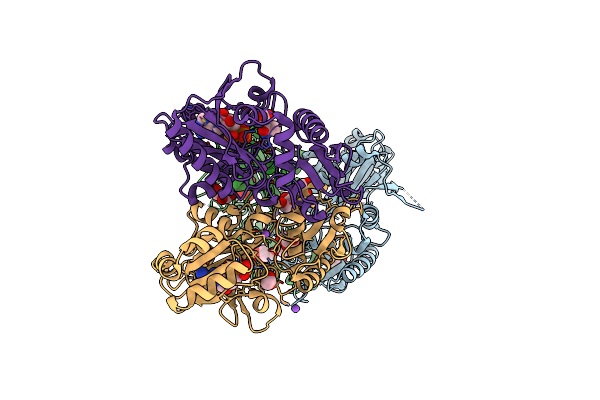
Organism: Escherichia coli (strain k12)
Method: X-RAY DIFFRACTION Resolution:1.80 Å Release Date: 2017-07-05 Classification: LYASE Ligands: ZN, CIT, PEG |
 |
Organism: Escherichia coli (strain k12)
Method: X-RAY DIFFRACTION Resolution:1.80 Å Release Date: 2017-07-05 Classification: LYASE Ligands: GOL, TRS, PEG, ZN |
 |
Organism: Escherichia coli (strain k12)
Method: X-RAY DIFFRACTION Resolution:2.00 Å Release Date: 2017-07-05 Classification: LYASE Ligands: GOL, ACT, ZN, PEG |
 |
Crystal Structure Of D-Alanine-D-Alanine Ligase From Acinetobacter Baumannii
Organism: Acinetobacter baumannii acicu
Method: X-RAY DIFFRACTION Resolution:2.19 Å Release Date: 2016-08-17 Classification: LIGASE |
 |
Crystal Structure Of D-Alanine-D-Alanine Ligase From Acinetobacter Baumannii, Space Group P212121
Organism: Acinetobacter baumannii acicu
Method: X-RAY DIFFRACTION Resolution:2.81 Å Release Date: 2016-08-17 Classification: LIGASE |
 |
Crystal Structure Of Adp Complexed D-Alanine-D-Alanine Ligase(Ddl) From Yersinia Pestis
Organism: Yersinia pestis
Method: X-RAY DIFFRACTION Resolution:2.28 Å Release Date: 2016-03-02 Classification: LIGASE |
 |
Crystal Structure Of Amp Complexed D-Alanine-D-Alanine Ligase(Ddl) From Yersinia Pestis
Organism: Yersinia pestis
Method: X-RAY DIFFRACTION Resolution:1.70 Å Release Date: 2016-03-02 Classification: LIGASE Ligands: AMP, NA, ACT, GOL |
 |
Crystal Structure Of Amp-Pnp Complexed D-Alanine-D-Alanine Ligase(Ddl) From Yersinia Pestis
Organism: Yersinia pestis
Method: X-RAY DIFFRACTION Resolution:2.50 Å Release Date: 2016-03-02 Classification: LIGASE Ligands: ANP, NA, MG |
 |
Crystal Structure Of Adp And D-Alanyl-D-Alanine Complexed D-Alanine-D-Alanine Ligase(Ddl) From Yersinia Pestis
Organism: Yersinia pestis
Method: X-RAY DIFFRACTION Resolution:2.40 Å Release Date: 2016-03-02 Classification: LIGASE |
 |
Crystal Structure Of Apo D-Alanine-D-Alanine Ligase(Ddl) From Yersinia Pestis
Organism: Yersinia pestis
Method: X-RAY DIFFRACTION Resolution:2.30 Å Release Date: 2016-01-13 Classification: LIGASE |
 |
Crystal Structure Of D-Alanine-D-Alnine Ligase From Xanthomonas Oryzae Pv. Oryzae With Amppnp
Organism: Xanthomonas oryzae pv. oryzae
Method: X-RAY DIFFRACTION Resolution:2.30 Å Release Date: 2014-02-19 Classification: LIGASE Ligands: ANP, MG |
 |
Crystal Structure Of D-Alanine-D-Alanine Ligase A From Xanthomonas Oryzae Pathovar Oryzae With Adp
Organism: Xanthomonas oryzae pv. oryzae
Method: X-RAY DIFFRACTION Resolution:2.10 Å Release Date: 2014-02-19 Classification: LIGASE Ligands: MG, ADP |