
Search Count: 5
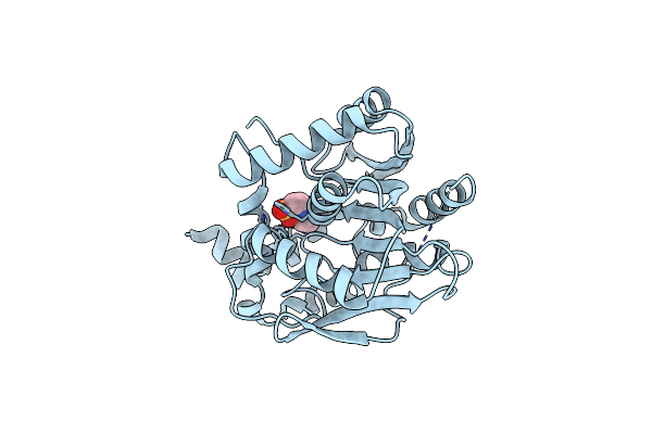 |
X-Ray Structure Of The Haloalkane Dehalogenase Halotag7 Circular Permutated At Positions 141-156 (Cphalotagdelta)
Organism: Rhodococcus sp.
Method: X-RAY DIFFRACTION Resolution:2.30 Å Release Date: 2023-10-11 Classification: HYDROLASE Ligands: NHE |
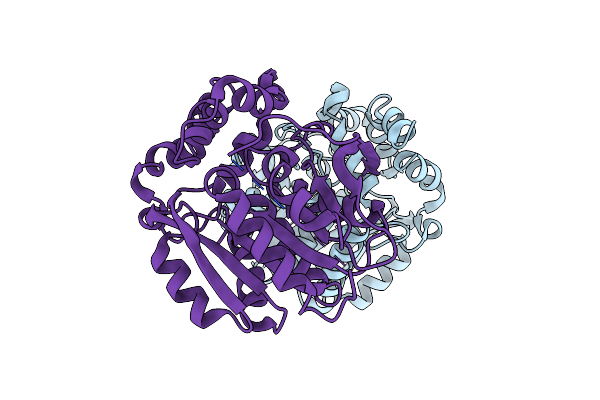 |
X-Ray Structure Of The Haloalkane Dehalogenase Halotag7 Circular Permutated At Positions 154-156 (Cphalotag7_154-156)
Organism: Rhodococcus sp.
Method: X-RAY DIFFRACTION Resolution:1.10 Å Release Date: 2023-10-11 Classification: HYDROLASE Ligands: CL |
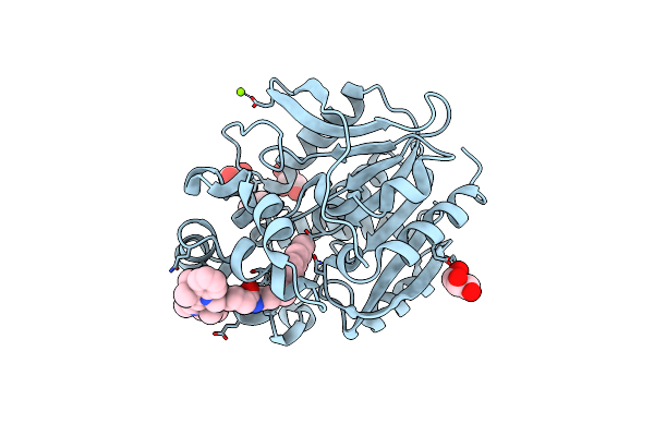 |
X-Ray Structure Of The Haloalkane Dehalogenase Halotag7 Labeled With A Chloroalkane Cyanine3 Fluorophore Substrate
Organism: Rhodococcus sp.
Method: X-RAY DIFFRACTION Resolution:1.50 Å Release Date: 2023-07-26 Classification: HYDROLASE Ligands: PJI, CL, GOL, MG |
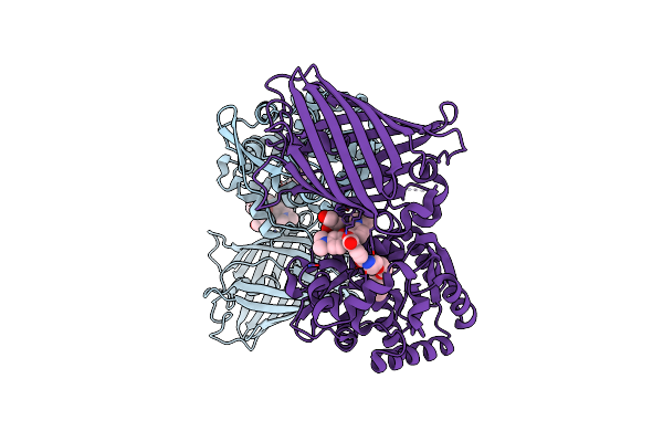 |
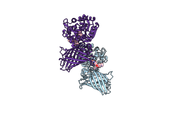
X-Ray Structure Of The Haloalkane Dehalogenase Halotag7 Fusion To The Green Fluorescent Protein Gfp (Chemog1) Labeled With A Chloroalkane Tetramethylrhodamine Fluorophore Substrate
Organism: Rhodococcus sp.
Method: X-RAY DIFFRACTION Resolution:1.80 Å Release Date: 2023-07-26 Classification: HYDROLASE Ligands: OEH, CL, GOL |
 |
X-Ray Structure Of The Interface Optimized Haloalkane Dehalogenase Halotag7 Fusion To The Green Fluorescent Protein Gfp (Chemog5-Tmr) Labeled With A Chloroalkane Tetramethylrhodamine Fluorophore Substrate
Organism: Rhodococcus sp.
Method: X-RAY DIFFRACTION Resolution:2.00 Å Release Date: 2023-07-26 Classification: HYDROLASE Ligands: OEH, CL |