
Search Count: 13
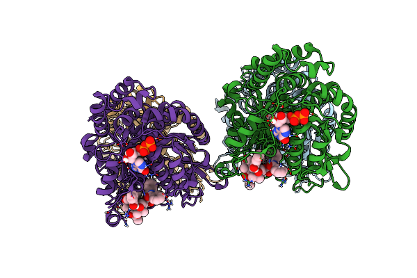 |
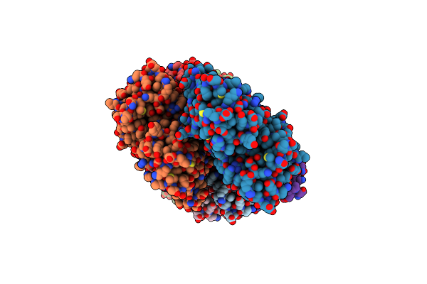
Low Curvature Lateral Interaction Within A 13-Protofilament, Taxol Stabilized Microtubule
Organism: Bos taurus
Method: ELECTRON MICROSCOPY Release Date: 2020-05-20 Classification: STRUCTURAL PROTEIN Ligands: GTP, MG, GDP, TA1 |
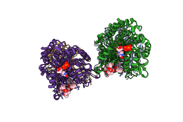 |
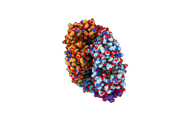
High Curvature Lateral Interaction Within A 13-Protofilament, Taxol Stabilized Microtubule
Organism: Bos taurus
Method: ELECTRON MICROSCOPY Release Date: 2020-05-20 Classification: STRUCTURAL PROTEIN Ligands: GTP, MG, GDP, TA1 |
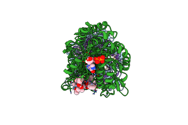 |
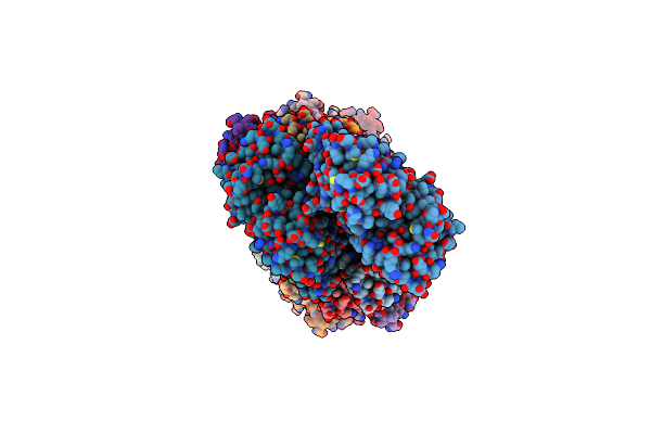
Organism: Bos taurus
Method: ELECTRON MICROSCOPY Release Date: 2020-05-20 Classification: STRUCTURAL PROTEIN Ligands: GTP, MG, GDP, TA1 |
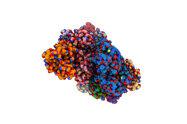 |
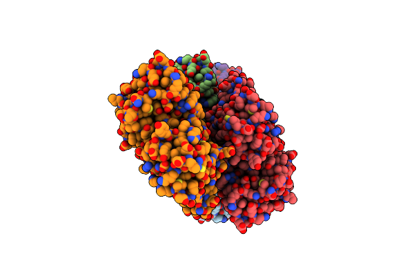
Organism: Homo sapiens, Oryctolagus cuniculus
Method: ELECTRON MICROSCOPY Release Date: 2020-01-08 Classification: STRUCTURAL PROTEIN Ligands: MG, ADP |
 |
Organism: Homo sapiens, Oryctolagus cuniculus
Method: ELECTRON MICROSCOPY Release Date: 2020-01-01 Classification: STRUCTURAL PROTEIN Ligands: MG, ADP |
 |
Organism: Homo sapiens, Oryctolagus cuniculus
Method: ELECTRON MICROSCOPY Release Date: 2020-01-01 Classification: STRUCTURAL PROTEIN Ligands: MG, ADP |
 |
Organism: Homo sapiens, Oryctolagus cuniculus
Method: ELECTRON MICROSCOPY Release Date: 2020-01-01 Classification: STRUCTURAL PROTEIN Ligands: MG, ADP |
 |
Organism: Oryctolagus cuniculus
Method: ELECTRON MICROSCOPY Release Date: 2020-01-01 Classification: STRUCTURAL PROTEIN Ligands: MG, ADP |
 |
Organism: Homo sapiens, Gallus gallus, Amanita phalloides
Method: ELECTRON MICROSCOPY Release Date: 2018-09-19 Classification: STRUCTURAL PROTEIN Ligands: ADP, MG |
 |
Crystal Structure Of Myosin 1B Residues 1-728 With Bound Sulfate And Calmodulin
Organism: Rattus norvegicus, Homo sapiens
Method: X-RAY DIFFRACTION Resolution:3.14 Å Release Date: 2018-02-28 Classification: MOTOR PROTEIN Ligands: SO4 |
 |
High-Resolution Cryo-Em Structures Of Actin-Bound Myosin States Reveal The Mechanism Of Myosin Force Sensing
Organism: Oryctolagus cuniculus, Rattus norvegicus, Unidentified, Amanita phalloides
Method: ELECTRON MICROSCOPY Release Date: 2018-01-31 Classification: STRUCTURAL PROTEIN Ligands: ADP, MG |
 |
High-Resolution Cryo-Em Structures Of Actin-Bound Myosin States Reveal The Mechanism Of Myosin Force Sensing
Organism: Rattus norvegicus, Unidentified, Oryctolagus cuniculus
Method: ELECTRON MICROSCOPY Release Date: 2018-01-31 Classification: STRUCTURAL PROTEIN Ligands: MG, ADP |
 |
High-Resolution Cryo-Em Structures Of Actin-Bound Myosin States Reveal The Mechanism Of Myosin Force Sensing
Organism: Oryctolagus cuniculus, Rattus norvegicus, Unidentified
Method: ELECTRON MICROSCOPY Release Date: 2018-01-31 Classification: STRUCTURAL PROTEIN Ligands: ADP, MG |