
Search Count: 28
 |
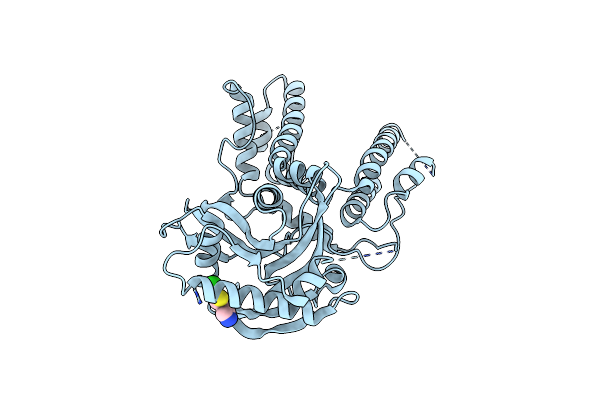
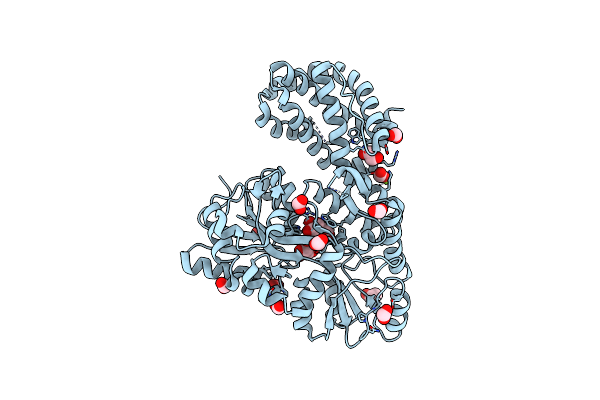
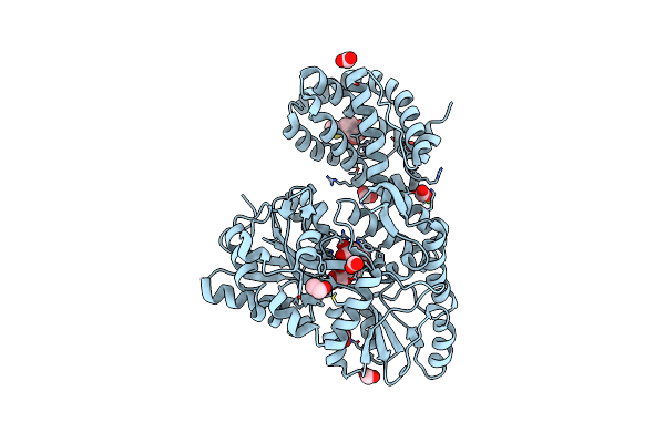
Crystal Structure Of Human Mbp-Myeloid Cell Leukemia 1 (Mcl-1) In Complex With Brd810 Inhibitor
Organism: Escherichia coli k-12, Homo sapiens
Method: X-RAY DIFFRACTION Resolution:1.56 Å Release Date: 2024-06-19 Classification: APOPTOSIS Ligands: MG, YI7, DMS, PGE |
 |
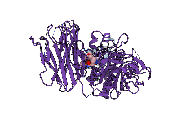
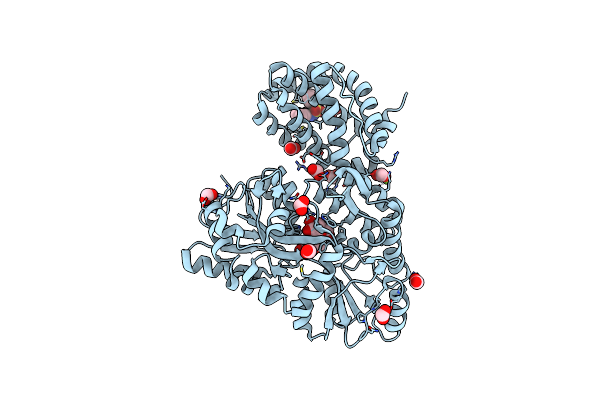
Organism: Homo sapiens
Method: X-RAY DIFFRACTION Resolution:1.53 Å Release Date: 2019-11-06 Classification: HYDROLASE/HYDROLASE Inhibitor Ligands: 063 |
 |
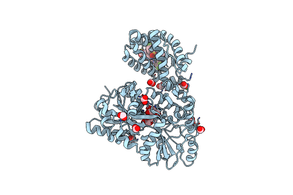
Organism: Homo sapiens
Method: X-RAY DIFFRACTION Resolution:2.15 Å Release Date: 2019-11-06 Classification: HYDROLASE Ligands: PV7 |
 |
Organism: Homo sapiens
Method: X-RAY DIFFRACTION Resolution:2.04 Å Release Date: 2019-11-06 Classification: HYDROLASE Ligands: EDO, PUV |
 |
Organism: Homo sapiens
Method: X-RAY DIFFRACTION Resolution:2.70 Å Release Date: 2019-11-06 Classification: HYDROLASE/Immune System Ligands: PVM |
 |
Organism: Homo sapiens
Method: X-RAY DIFFRACTION Resolution:2.73 Å Release Date: 2019-11-06 Classification: HYDROLASE/Immune System Ligands: PVJ |
 |
Organism: Homo sapiens
Method: X-RAY DIFFRACTION Resolution:2.64 Å Release Date: 2019-11-06 Classification: HYDROLASE Ligands: PVV |
 |
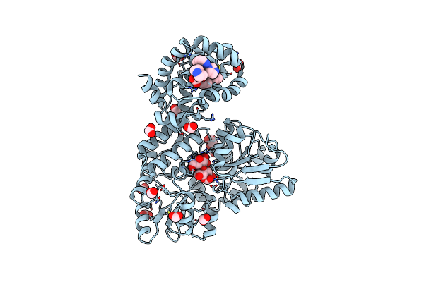
Crystal Structure Of Polymerase Acid Protein (Pa) From Influenza A Virus, Wilson-Smith/1933 (H1N1) Bound To Follow On Fragment Ebsi-4723 4-(5-Chlorothiophen-2-Yl)-1H-Pyrazole
Organism: Influenza a virus (strain a/wilson-smith/1933 h1n1)
Method: X-RAY DIFFRACTION Resolution:2.45 Å Release Date: 2017-03-08 Classification: TRANSCRIPTION Ligands: 8QG |
 |
Organism: Homo sapiens
Method: X-RAY DIFFRACTION Resolution:1.70 Å Release Date: 2015-05-06 Classification: Apoptosis/Inhibitor Ligands: ZN, 865, POP |
 |
Organism: Escherichia coli, Homo sapiens
Method: X-RAY DIFFRACTION Resolution:1.90 Å Release Date: 2015-05-06 Classification: APOPTOSIS Ligands: MG, EDO, FMT |
 |
Organism: Escherichia coli, Homo sapiens
Method: X-RAY DIFFRACTION Resolution:2.35 Å Release Date: 2015-05-06 Classification: APOPTOSIS Ligands: 865, EDO, FMT |
 |
Organism: Escherichia coli, Homo sapiens
Method: X-RAY DIFFRACTION Resolution:1.55 Å Release Date: 2015-05-06 Classification: APOPTOSIS Ligands: 19H, MG, FMT, NA, EDO |
 |
Organism: Escherichia coli, Homo sapiens
Method: X-RAY DIFFRACTION Resolution:2.40 Å Release Date: 2015-05-06 Classification: APOPTOSIS Ligands: CL, MG, 3R4 |
 |
Organism: Escherichia coli, Homo sapiens
Method: X-RAY DIFFRACTION Resolution:1.90 Å Release Date: 2015-05-06 Classification: APOPTOSIS Ligands: MG, EDO, FMT, 3R6 |
 |
Organism: Escherichia coli, Homo sapiens
Method: X-RAY DIFFRACTION Resolution:2.00 Å Release Date: 2015-05-06 Classification: APOPTOSIS Ligands: 3R7, FMT, MG |
 |
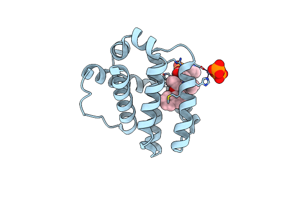
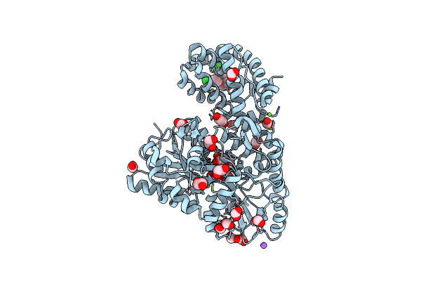
A Single Diastereomer Of A Macrolactam Core Binds Specifically To Myeloid Cell Leukemia 1 (Mcl1)
Organism: Escherichia coli o157:h7, Homo sapiens
Method: X-RAY DIFFRACTION Resolution:1.85 Å Release Date: 2014-11-19 Classification: apoptosis/inhibitor Ligands: 3M6, MG, FMT |
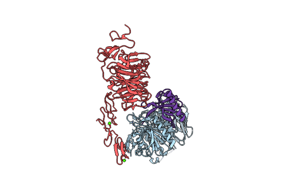 |
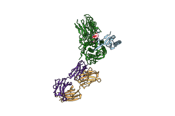
The Structure Of The Ldlr/Pcsk9 Complex Reveals The Receptor In An Extended Conformation
Organism: Homo sapiens
Method: X-RAY DIFFRACTION Resolution:3.30 Å Release Date: 2011-10-26 Classification: HYDROLASE/LIPID BINDING PROTEIN Ligands: CA |
 |
The Structure Of The Ldlr/Pcsk9 Complex Reveals The Receptor In An Extended Conformation
Organism: Homo sapiens
Method: X-RAY DIFFRACTION Resolution:4.20 Å Release Date: 2011-10-26 Classification: HYDROLASE/LIPID BINDING PROTEIN Ligands: CA |
 |
Organism: Homo sapiens
Method: X-RAY DIFFRACTION Resolution:2.70 Å Release Date: 2010-11-03 Classification: HYDROLASE/ANTIBODY Ligands: CA |
 |
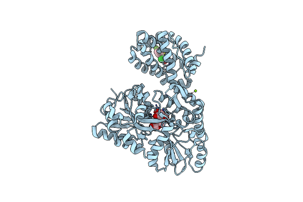
Crystal Structure Of L-Rhamnonate Dehydratase From Salmonella Typhimurium Complexed With Mg And 3-Deoxy-L-Rhamnonate
Organism: Salmonella typhimurium lt2
Method: X-RAY DIFFRACTION Resolution:2.00 Å Release Date: 2008-09-23 Classification: LYASE Ligands: 3LR, MG, 1N5 |