
Search Count: 21
 |
Organism: Vaccinia virus western reserve
Method: ELECTRON MICROSCOPY Release Date: 2025-09-03 Classification: VIRAL PROTEIN Ligands: NAG |
 |
Organism: Vaccinia virus western reserve
Method: X-RAY DIFFRACTION Release Date: 2025-09-03 Classification: VIRAL PROTEIN Ligands: PEG |
 |
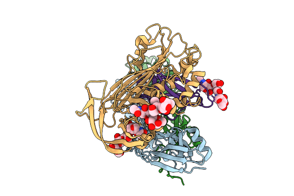
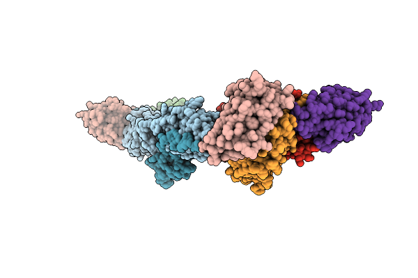
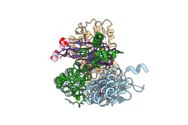
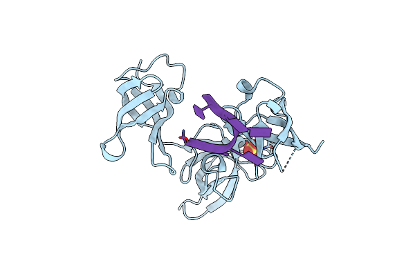
Structure Of A16/G9 (Vaccinia Virus) In Complex With Vhh D07, Vhh B01 And Vhh C05
Organism: Vaccinia virus western reserve, Vicugna pacos
Method: X-RAY DIFFRACTION Release Date: 2025-09-03 Classification: VIRAL PROTEIN Ligands: K, PO4 |
 |
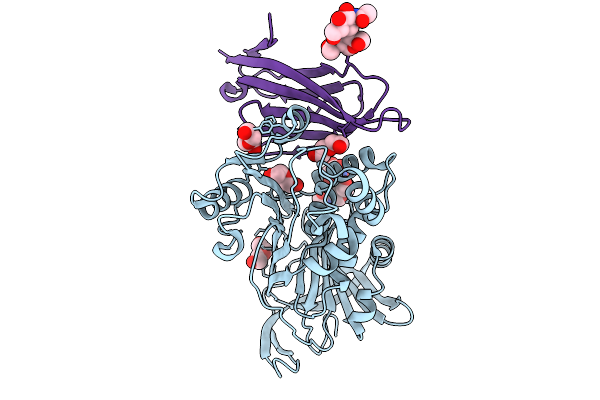
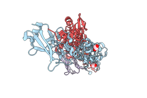
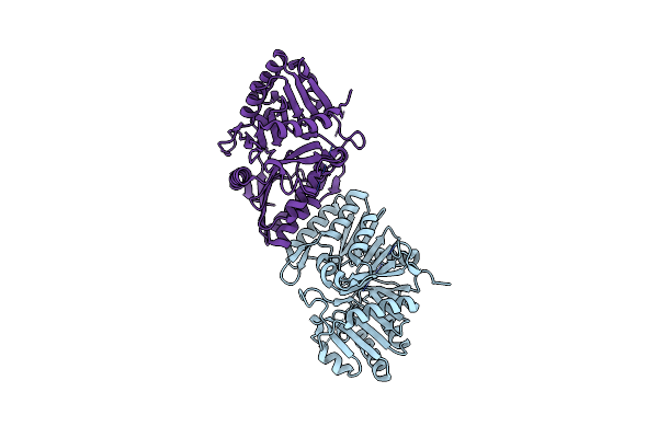
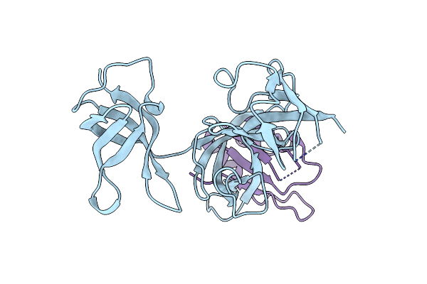
Structure Of A16/G9 (G9 Mutant - H44Y) Of Vaccinia Virus In Complex With Vhh D07
Organism: Vaccinia virus western reserve, Vicugna pacos
Method: X-RAY DIFFRACTION Release Date: 2025-09-03 Classification: VIRAL PROTEIN |
 |
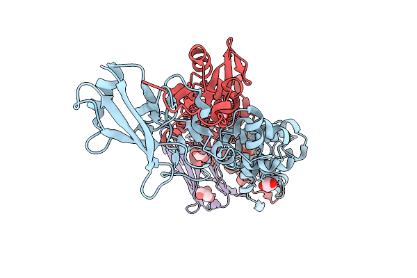
Organism: Vaccinia virus western reserve, Vicugna pacos
Method: X-RAY DIFFRACTION Release Date: 2025-09-03 Classification: VIRAL PROTEIN Ligands: PO4, SO4, PEG |
 |
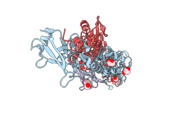
Organism: Vaccinia virus western reserve, Vicugna pacos
Method: X-RAY DIFFRACTION Release Date: 2025-09-03 Classification: VIRAL PROTEIN Ligands: MES, GOL |
 |
Organism: Vaccinia virus western reserve, Vicugna pacos
Method: X-RAY DIFFRACTION Release Date: 2025-09-03 Classification: VIRAL PROTEIN Ligands: MES, GOL |
 |
Organism: Vaccinia virus western reserve, Vicugna pacos
Method: X-RAY DIFFRACTION Release Date: 2025-09-03 Classification: VIRAL PROTEIN Ligands: GOL, MES |
 |
Organism: Vaccinia virus western reserve
Method: ELECTRON MICROSCOPY Release Date: 2025-09-03 Classification: VIRAL PROTEIN Ligands: NAG |
 |
Organism: Vaccinia virus western reserve
Method: X-RAY DIFFRACTION Resolution:2.10 Å Release Date: 2025-03-12 Classification: VIRAL PROTEIN |
 |
Organism: Vaccinia virus western reserve
Method: X-RAY DIFFRACTION Resolution:2.82 Å Release Date: 2025-03-12 Classification: VIRAL PROTEIN Ligands: CIT, GOL |
 |
Organism: Vaccinia virus western reserve
Method: X-RAY DIFFRACTION Resolution:2.60 Å Release Date: 2025-03-12 Classification: VIRAL PROTEIN Ligands: CIT, GOL |
 |
Organism: Vaccinia virus western reserve
Method: X-RAY DIFFRACTION Resolution:4.00 Å Release Date: 2025-03-12 Classification: VIRAL PROTEIN Ligands: GOL |
 |
Organism: Vaccinia virus western reserve
Method: X-RAY DIFFRACTION Resolution:3.80 Å Release Date: 2025-03-12 Classification: VIRAL PROTEIN Ligands: CIT, GOL, A1IDM |
 |
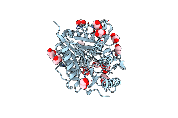
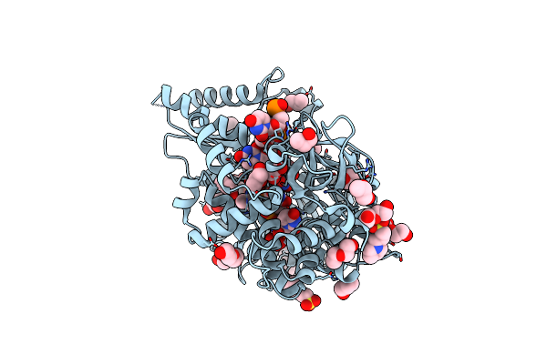
Structure Of The F13 Protein (A295E Mutant) Of Vaccinia Virus In Complex With Tecovirimat
Organism: Vaccinia virus western reserve
Method: X-RAY DIFFRACTION Resolution:3.50 Å Release Date: 2025-03-12 Classification: VIRAL PROTEIN Ligands: GOL, A1ICJ |
 |
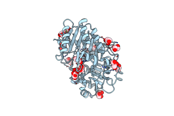
Organism: Vaccinia virus western reserve
Method: X-RAY DIFFRACTION Resolution:2.60 Å Release Date: 2025-03-12 Classification: VIRAL PROTEIN Ligands: A1ICJ, CIT, GOL |
 |
Organism: Drosophila melanogaster
Method: X-RAY DIFFRACTION Resolution:1.60 Å Release Date: 2022-12-07 Classification: RNA BINDING PROTEIN Ligands: SO4 |
 |
Organism: Drosophila melanogaster
Method: X-RAY DIFFRACTION Resolution:2.99 Å Release Date: 2022-12-07 Classification: RNA BINDING PROTEIN |
 |
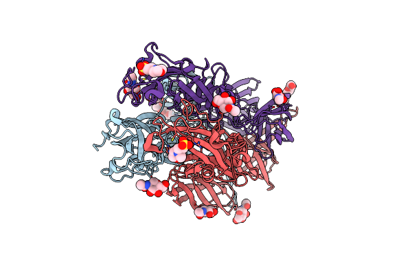
Crystal Structure Of The Deah-Box Atpase Prp2 In Complex With Adp-Bef3 And Ssrna
Organism: Chaetomium thermophilum (strain dsm 1495 / cbs 144.50 / imi 039719), Synthetic construct
Method: X-RAY DIFFRACTION Resolution:2.10 Å Release Date: 2021-04-21 Classification: HYDROLASE Ligands: BEF, ADP, MG, PEG, GOL, MPO, CL, PGO, HEZ, POL, EDO |
 |
Structure Of Rvfv Envelope Protein Gc In Postfusion Conformation In Complex With Mes
Organism: Rift valley fever virus
Method: X-RAY DIFFRACTION Resolution:2.50 Å Release Date: 2017-11-01 Classification: VIRAL PROTEIN Ligands: NAG, MES |