
Search Count: 26
 |
Organism: Homo sapiens
Method: SOLUTION NMR Release Date: 2015-02-11 Classification: LIGASE Ligands: ZN |
 |
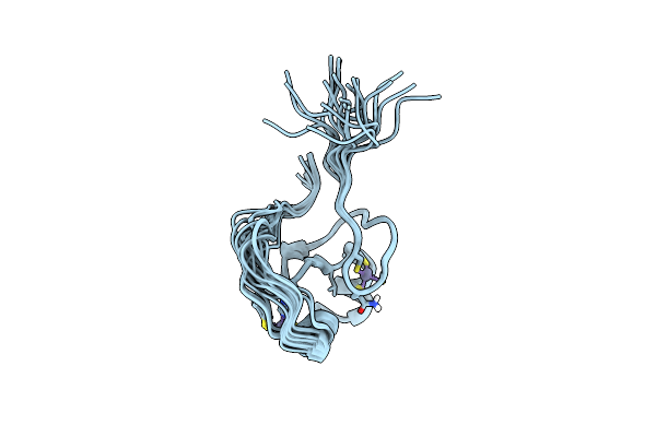
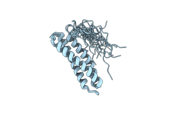
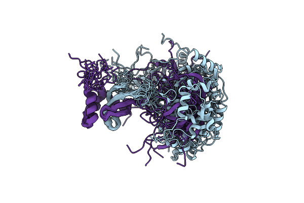
Solution Structure Of The Trim19 B-Box1 (B1) Of Human Promyelocytic Leukemia (Pml)
Organism: Homo sapiens
Method: SOLUTION NMR Release Date: 2014-11-12 Classification: METAL BINDING PROTEIN Ligands: ZN |
 |
Organism: Homo sapiens
Method: X-RAY DIFFRACTION Resolution:1.63 Å Release Date: 2014-10-15 Classification: PROTEIN TRANSPORT |
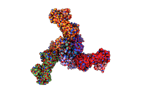 |
Crystal Structure Of Ternary Complex Of Trail, Dr5, And Fab Fragment From A Dr5 Agonist Antibody
Organism: Homo sapiens
Method: X-RAY DIFFRACTION Resolution:3.30 Å Release Date: 2014-09-03 Classification: Apoptosis/Immune System Ligands: ZN |
 |
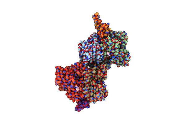
Three-Dimensional Structure Of The C-Terminal Dna-Binding Domain Of Rsta Protein From Klebsiella Pneumoniae
Organism: Klebsiella pneumoniae
Method: SOLUTION NMR Release Date: 2014-07-16 Classification: SIGNALING PROTEIN |
 |
Crystal Structure Of Klebsiella Pneumoniae Rsta Dna-Binding Domain In Complex With Rsta Box
Organism: Klebsiella pneumoniae
Method: X-RAY DIFFRACTION Resolution:2.70 Å Release Date: 2014-07-16 Classification: TRANSCRIPTION REGULATOR/DNA |
 |
Crystal Structure Of Klebsiella Pneumoniae Rsta Bef3-Activated N-Terminal Receiver Domain
Organism: Klebsiella pneumoniae
Method: X-RAY DIFFRACTION Resolution:3.18 Å Release Date: 2014-07-16 Classification: TRANSCRIPTION REGULATOR Ligands: MG, BEF |
 |
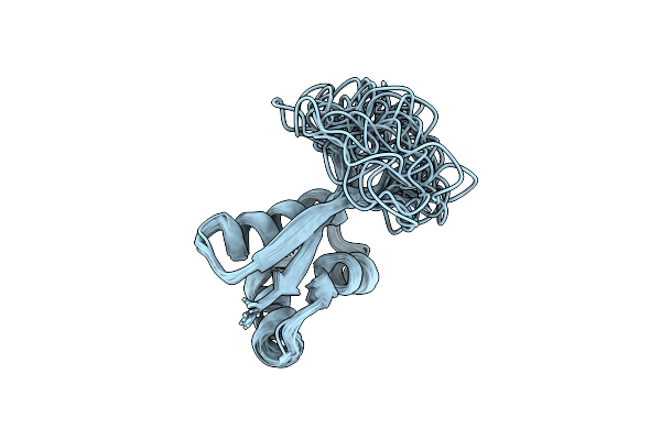
Structure Of The N-Terminal Domian Of Human Coronavirus Oc43 Nucleocapsid Protein
Organism: Human coronavirus
Method: X-RAY DIFFRACTION Resolution:2.00 Å Release Date: 2013-05-08 Classification: RNA BINDING PROTEIN Ligands: TRS |
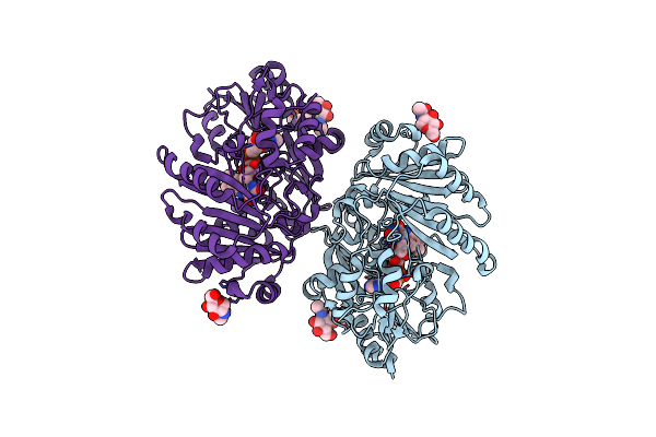 |
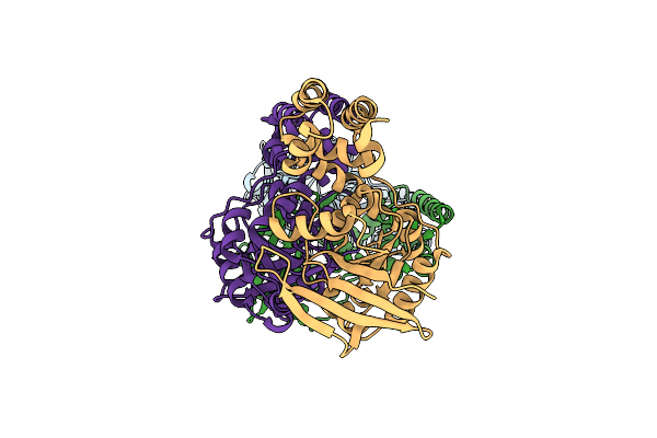
Crystal Structure Of Bermuda Grass Isoallergen Bg60 Provides Insight Into The Various Cross-Allergenicity Of The Pollen Group 4 Allergens
Organism: Cynodon dactylon
Method: X-RAY DIFFRACTION Resolution:2.15 Å Release Date: 2012-12-05 Classification: OXIDOREDUCTASE Ligands: FAD, NAG |
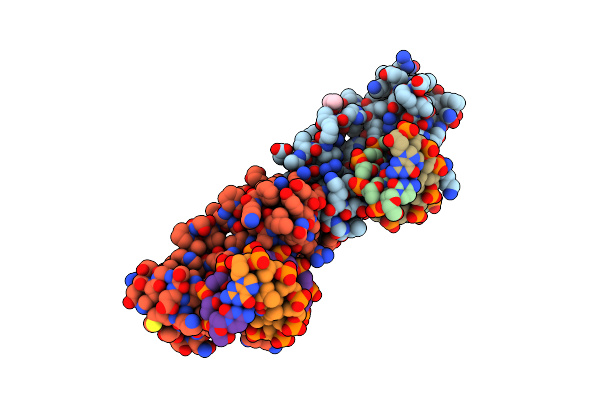 |
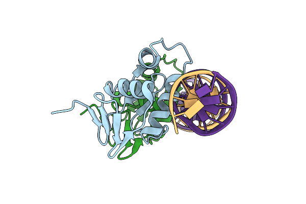
The Structure Of Protozoan Parasite Trichomonas Vaginalis Myb2 In Complex With Mre-2F-13 Dna
Organism: Trichomonas vaginalis
Method: X-RAY DIFFRACTION Resolution:2.03 Å Release Date: 2011-08-03 Classification: transcription/DNA Ligands: IPA |
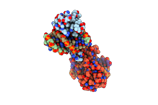 |
The Structure Of Protozoan Parasite Trichomonas Vaginalis Myb2 In Complex With Mre-1-12 Dna
Organism: Trichomonas vaginalis
Method: X-RAY DIFFRACTION Resolution:2.00 Å Release Date: 2011-08-03 Classification: transcription/DNA |
 |
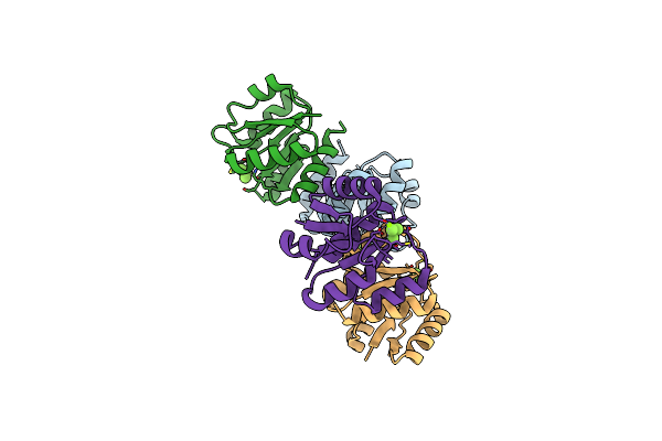
The Crystal Structure Of The Subunit Binding Of Human Dihydrolipoamide Transacylase (E2B) Bound To Human Dihydrolipoamide Dehydrogenase (E3)
Organism: Homo sapiens
Method: X-RAY DIFFRACTION Resolution:2.40 Å Release Date: 2011-05-04 Classification: OXIDOREDUCTASE/PROTEIN BINDING Ligands: BME, FAD, NHE |
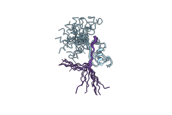 |
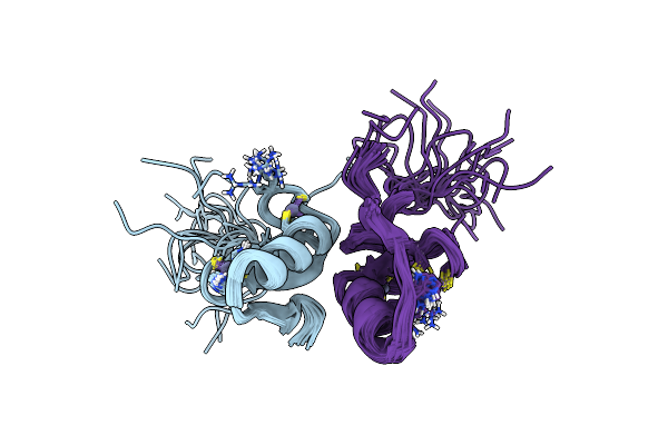
Phosphorylation Of Sumo-Interacting Motif By Ck2 Enhances Daxx Sumo Binding Activity
Organism: Homo sapiens
Method: SOLUTION NMR Release Date: 2010-12-01 Classification: TRANSCRIPTION, APOPTOSIS |
 |
Organism: Pyrococcus furiosus
Method: X-RAY DIFFRACTION Resolution:2.70 Å Release Date: 2010-05-26 Classification: METAL TRANSPORT |
 |
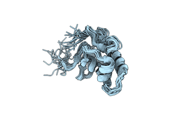
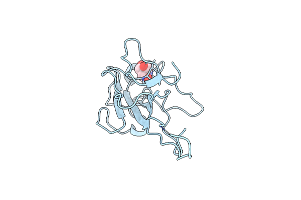
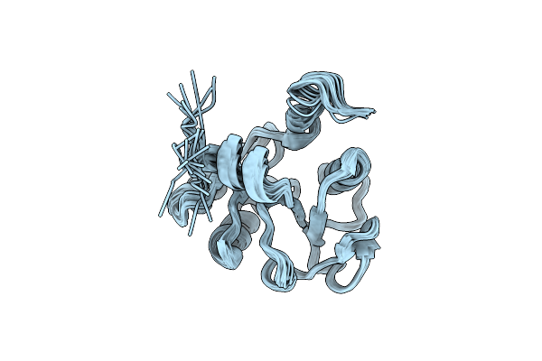
Solution Structure Of Putative Ferrous Iron Transport Protein C (Feoc) Of Klebsiella Pneumoniae
Organism: Klebsiella pneumoniae subsp. pneumoniae
Method: SOLUTION NMR Release Date: 2009-01-27 Classification: METAL BINDING PROTEIN |
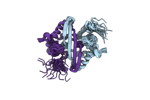 |
Solution Structure Of Stereo-Array Isotope Labelled (Sail) C-Terminal Dimerization Domain Of Sars Coronavirus Nucleocapsid Protein
Organism: Human sars coronavirus
Method: SOLUTION NMR Release Date: 2008-08-26 Classification: VIRAL PROTEIN |
 |
Nmr Solution Structure Of Blo T 5, A Major Mite Allergen From Blomia Tropicalis
|
 |
Domain-Swapped Dimer Of The Pwwp Module Of Human Hepatoma-Derived Growth Factor
Organism: Homo sapiens
Method: SOLUTION NMR Release Date: 2007-09-04 Classification: HORMONE/GROWTH FACTOR |
 |
Solution Structure Of Putative Coa-Binding Protein Of Klebsiella Pneumoniae
Organism: Klebsiella pneumoniae
Method: SOLUTION NMR Release Date: 2007-08-14 Classification: LIGAND BINDING PROTEIN |
 |
Crystal Structure Of Oligomerization Domain Of Sars Coronavirus Nucleocapsid Protein.
Organism: Sars coronavirus
Method: X-RAY DIFFRACTION Resolution:2.50 Å Release Date: 2007-04-10 Classification: VIRAL PROTEIN |