
Search Count: 68
 |
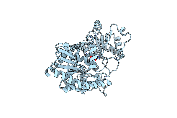
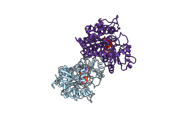
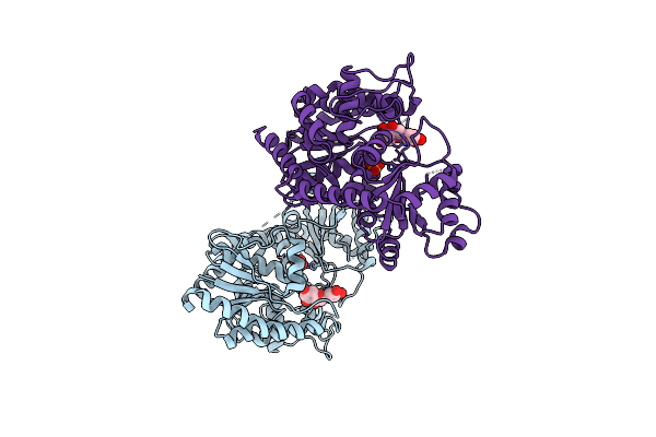
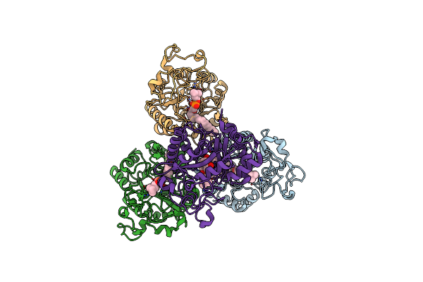
Cryo-Em Structure Of Tmprss2 In Complex With Fab Fragments Of 752 Mab And 2228 Mab
Organism: Homo sapiens, Mus musculus
Method: ELECTRON MICROSCOPY Release Date: 2025-10-15 Classification: MEMBRANE PROTEIN |
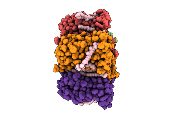 |
Cryo-Em Structure Of The Zeaxanthin-Bound Light-Driven Proton Pumping Rhodopsin, Nm-R1
Organism: Nonlabens marinus s1-08
Method: ELECTRON MICROSCOPY Release Date: 2025-07-30 Classification: MEMBRANE PROTEIN Ligands: RET, R16, D12, K3I |
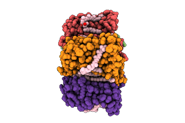 |
Cryo-Em Structure Of The Myxol-Bound Light-Driven Proton Pumping Rhodopsin, Nm-R1
Organism: Nonlabens marinus s1-08
Method: ELECTRON MICROSCOPY Release Date: 2025-07-30 Classification: MEMBRANE PROTEIN Ligands: RET, R16, D12, A1L4O |
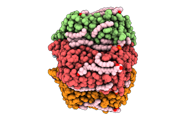 |
Cryo-Em Structure Of The Myxol-Bound Light-Driven Chloride Ion-Pumping Rhodopsin, Nm-R3
Organism: Nonlabens marinus s1-08
Method: ELECTRON MICROSCOPY Release Date: 2025-07-30 Classification: MEMBRANE PROTEIN Ligands: RET, A1L4O, CL, PC1, PLC, R16, 8K6, D12, C14, D10 |
 |
Cryo-Em Structure Of The Light-Driven Chloride Ion-Pumping Rhodopsin, Nm-R3
Organism: Nonlabens marinus s1-08
Method: ELECTRON MICROSCOPY Release Date: 2025-07-30 Classification: MEMBRANE PROTEIN Ligands: RET, CL, PC1, PLC, D12, R16, 8K6, C14 |
 |
Organism: Chiba virus, Homo sapiens
Method: ELECTRON MICROSCOPY Release Date: 2024-04-17 Classification: VIRUS |
 |
Structure Of Glycerophosphoethanolamine Ethanolaminephosphodiesterase From Streptomyces Sanglieri
Organism: Streptomyces sanglieri
Method: X-RAY DIFFRACTION Resolution:2.10 Å Release Date: 2024-04-03 Classification: HYDROLASE Ligands: CA, GOL |
 |
Structure Of Flavone 4'-O-Glucoside 7-O-Glucosyltransferase From Nemophila Menziesii, Apo Form
Organism: Nemophila menziesii
Method: X-RAY DIFFRACTION Resolution:2.40 Å Release Date: 2024-02-14 Classification: PLANT PROTEIN Ligands: SO4 |
 |
Structure Of Flavone 4'-O-Glucoside 7-O-Glucosyltransferase From Nemophila Menziesii, Complex With Udp-Glucose
Organism: Nemophila menziesii
Method: X-RAY DIFFRACTION Resolution:2.10 Å Release Date: 2024-02-14 Classification: PLANT PROTEIN Ligands: UPG |
 |
Structure Of Flavone 4'-O-Glucoside 7-O-Glucosyltransferase From Nemophila Menziesii, Complex With Luteolin
Organism: Nemophila menziesii
Method: X-RAY DIFFRACTION Resolution:2.43 Å Release Date: 2024-02-14 Classification: PLANT PROTEIN Ligands: LU2, SO4 |
 |
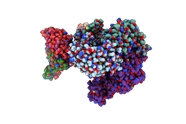
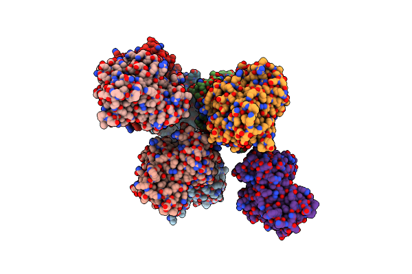
Crystal Structure Of P Domain From Norovirus Gi.4 Capsid Protein In Complex With Broad Specificity Antibody Single-Chain Fv Fragment Cv-2F5.
Organism: Chiba virus, Homo sapiens
Method: X-RAY DIFFRACTION Resolution:2.70 Å Release Date: 2024-01-31 Classification: VIRAL PROTEIN |
 |
The Crystal Structure Of Cyanorhodopsin-Ii (Cyr-Ii) P7104R From Nodosilinea Nodulosa Pcc 7104
Organism: Nodosilinea nodulosa pcc 7104
Method: X-RAY DIFFRACTION Resolution:2.07 Å Release Date: 2023-10-25 Classification: MEMBRANE PROTEIN Ligands: RET, PG4, HEX, OCT, C14, R16, SO4, CL |
 |
Organism: Thermocrispum sp. rd004668
Method: X-RAY DIFFRACTION Resolution:2.57 Å Release Date: 2023-02-08 Classification: HYDROLASE |
 |
Organism: Thermocrispum sp. rd004668
Method: X-RAY DIFFRACTION Resolution:2.29 Å Release Date: 2023-02-08 Classification: HYDROLASE |
 |
Complex Structure Of Lysoplasmalogen Specific Phopholipase D, F211L Mutant With Lpc
Organism: Thermocrispum sp. rd004668
Method: X-RAY DIFFRACTION Resolution:2.69 Å Release Date: 2023-02-08 Classification: HYDROLASE Ligands: KIP |
 |
Organism: Thermocrispum sp. rd004668
Method: X-RAY DIFFRACTION Resolution:2.91 Å Release Date: 2023-01-04 Classification: HYDROLASE Ligands: CA |
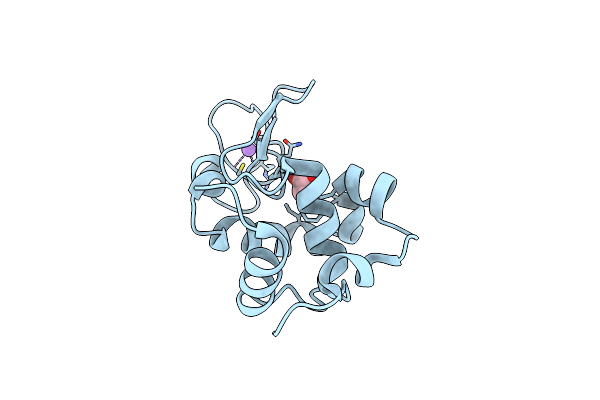 |
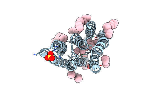
Crystal Structure Of Hen Egg White Lysozyme Introduced With O-(2-Nitrobenzyl)-L-Tyrosine
Organism: Gallus gallus
Method: X-RAY DIFFRACTION Resolution:1.44 Å Release Date: 2022-09-21 Classification: BIOSYNTHETIC PROTEIN |
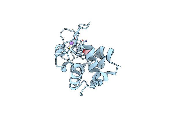 |
Crystal Structure Of Photolysed Hen Egg White Lysozyme Introduced With O-(2-Nitrobenzyl)-L-Tyrosine
Organism: Gallus gallus
Method: X-RAY DIFFRACTION Resolution:1.39 Å Release Date: 2022-09-21 Classification: BIOSYNTHETIC PROTEIN |
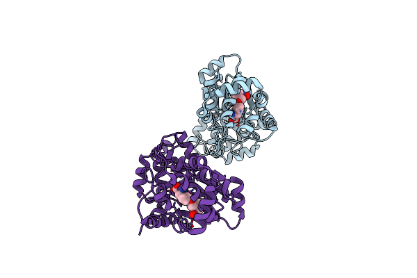 |
Crystal Structure Of O-(2-Nitrobenzyl)-L-Tyrosine-Trna Sythetase In Complex With O-(2-Nitrobenzyl)-L-Tyrosine
Organism: Methanocaldococcus jannaschii
Method: X-RAY DIFFRACTION Resolution:2.79 Å Release Date: 2022-09-21 Classification: TRANSCRIPTION Ligands: J2F |
 |
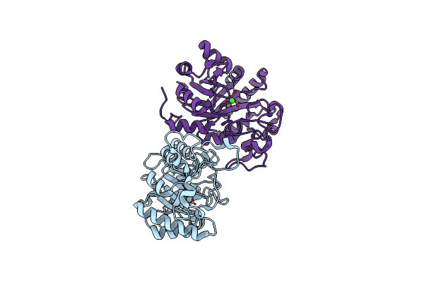
Complex Structure Of Serine Hydroxymethyltransferase From Enterococcus Faecium And Its Inhibitor
Organism: Enterococcus faecium
Method: X-RAY DIFFRACTION Resolution:2.28 Å Release Date: 2022-07-06 Classification: TRANSFERASE Ligands: 5M5, PLP, CL |