
Search Count: 14
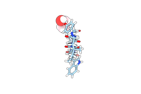 |
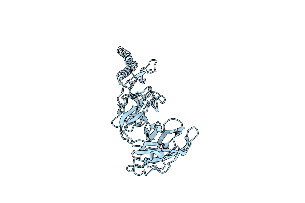
Structure Of The Amyloid-Forming Peptide Lyiqwl From Tc5B, Grown From 30% Ethanol
Organism: Synthetic construct
Method: X-RAY DIFFRACTION Resolution:1.50 Å Release Date: 2023-08-02 Classification: PROTEIN FIBRIL Ligands: EOH |
 |
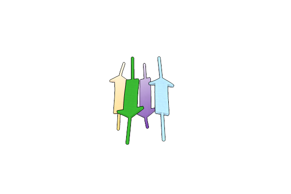
Structure Of The Amyloid-Forming Peptide Lyiqwl From Tc5B, Grown From 30% Acetonitrile
Organism: Synthetic construct
Method: X-RAY DIFFRACTION Resolution:1.25 Å Release Date: 2023-08-02 Classification: PROTEIN FIBRIL Ligands: TFA, CCN |
 |
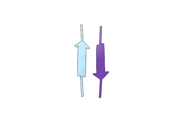
Structure Of The Amyloid-Forming Peptide Lyiqwl From Tc5B, Grown From 10% Ethanol
Organism: Synthetic construct
Method: X-RAY DIFFRACTION Resolution:1.35 Å Release Date: 2023-08-02 Classification: PROTEIN FIBRIL Ligands: TFA, EOH |
 |
Organism: Homo sapiens
Method: X-RAY DIFFRACTION Resolution:1.55 Å Release Date: 2023-08-02 Classification: PROTEIN FIBRIL Ligands: HOH |
 |
Organism: Synthetic construct
Method: X-RAY DIFFRACTION Resolution:1.30 Å Release Date: 2023-08-02 Classification: PROTEIN FIBRIL |
 |
Structure Of The Amyloid-Forming Peptide Lfiewl From Exendin-4, Grown From Water
Organism: Heloderma suspectum
Method: X-RAY DIFFRACTION Resolution:1.75 Å Release Date: 2023-08-02 Classification: PROTEIN FIBRIL |
 |
Structure Of The Amyloid-Forming Peptide Lyiqwl From Tc5B, Grown From Water
Organism: Synthetic construct
Method: X-RAY DIFFRACTION Resolution:0.90 Å Release Date: 2023-08-02 Classification: PROTEIN FIBRIL |
 |
Structure Of The Amyloid-Forming Peptide Lfiewl From Exendin-4, Grown From Acetonitrile / Water
Organism: Heloderma suspectum
Method: X-RAY DIFFRACTION Resolution:1.24 Å Release Date: 2023-08-02 Classification: PROTEIN FIBRIL |
 |
Organism: Synthetic construct
Method: X-RAY DIFFRACTION Resolution:1.50 Å Release Date: 2023-08-02 Classification: PROTEIN FIBRIL |
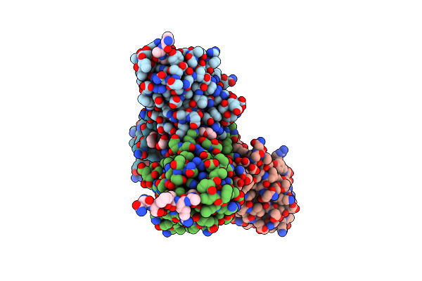 |
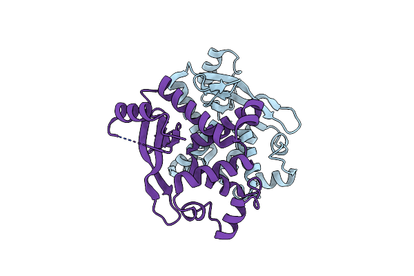
Crystal Structure Of Neuropilin-1 B1 Domain In Complex With Sars-Cov-2 S1 C-End Rule (Cendr) Peptide
Organism: Homo sapiens, Severe acute respiratory syndrome coronavirus 2
Method: X-RAY DIFFRACTION Resolution:2.36 Å Release Date: 2020-10-28 Classification: SIGNALING PROTEIN Ligands: PEG, GOL |
 |
Organism: Salmonella typhimurium
Method: ELECTRON MICROSCOPY Release Date: 2020-02-12 Classification: MOTOR PROTEIN |
 |
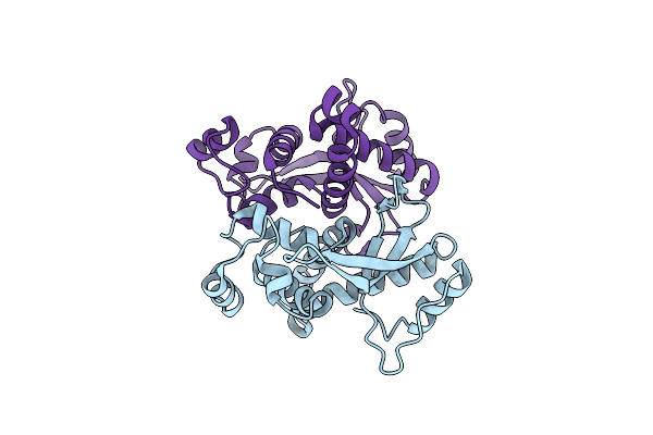
Structure Of Salmonella Flagellar Hook Reveals Intermolecular Domain Interactions For The Universal Joint Function
Organism: Salmonella enterica subsp. enterica serovar typhimurium
Method: ELECTRON MICROSCOPY Release Date: 2019-10-16 Classification: MOTOR PROTEIN |
 |
Organism: Streptococcus phage dt1
Method: X-RAY DIFFRACTION Resolution:1.96 Å Release Date: 2018-06-06 Classification: VIRAL PROTEIN |
 |
Organism: Streptococcus phage 73
Method: X-RAY DIFFRACTION Resolution:2.50 Å Release Date: 2018-06-06 Classification: VIRAL PROTEIN |