
Search Count: 210
 |
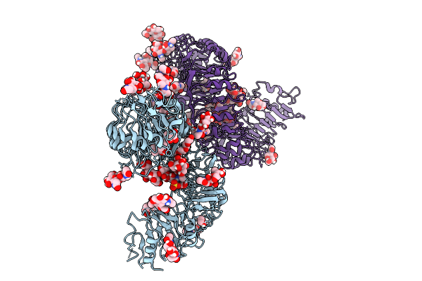
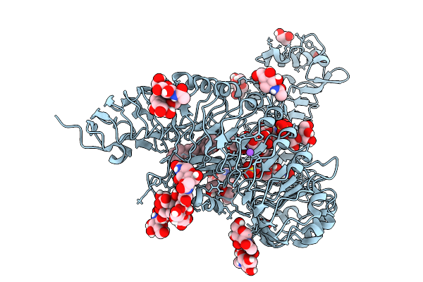
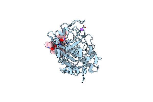
Structure Of Cathepsin B1 From Schistosoma Mansoni (Smcb1) In Complex With A Carborane Inhibitor
Organism: Schistosoma mansoni
Method: X-RAY DIFFRACTION Release Date: 2025-10-29 Classification: HYDROLASE Ligands: A1IHQ, EDO |
 |
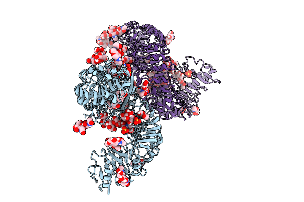
Crystal Structure Of The Brl3 Ectodomain From Arabidopsis Thaliana In Complex With Brassinolide.
Organism: Arabidopsis thaliana
Method: X-RAY DIFFRACTION Release Date: 2025-09-10 Classification: MEMBRANE PROTEIN Ligands: BLD, NAG, 1PE, EDO, SO4, GLY, ACT |
 |
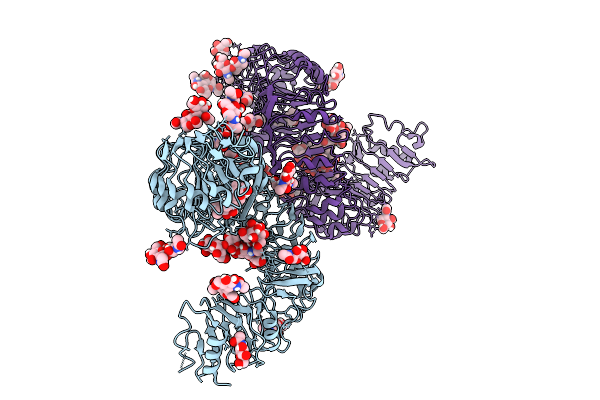
Crystal Structure Of The Bri1 Ectodomain From Arabidopsis Thaliana In Complex With Castasterone.
Organism: Arabidopsis thaliana
Method: X-RAY DIFFRACTION Release Date: 2025-09-10 Classification: MEMBRANE PROTEIN Ligands: SO4, EDO, NAG, CIT, A1JME |
 |
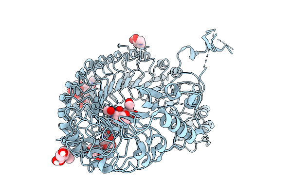
Crystal Structure Of The Bri1 Ectodomain From Arabidopsis Thaliana In Complex With Typhasterol.
Organism: Arabidopsis thaliana
Method: X-RAY DIFFRACTION Release Date: 2025-09-10 Classification: MEMBRANE PROTEIN Ligands: A1JMF, SO4, EDO, NAG, CIT |
 |
Crystal Structure Of The Bri1 Ectodomain From Arabidopsis Thaliana In Complex With 24-Epibrassinolide.
Organism: Arabidopsis thaliana
Method: X-RAY DIFFRACTION Release Date: 2025-09-10 Classification: MEMBRANE PROTEIN Ligands: A1JMG, EDO, NAG |
 |
Crystal Structure Of The Bri1 Ectodomain From Arabidopsis Thaliana In Complex With 28-Homobrassinolide.
Organism: Arabidopsis thaliana
Method: X-RAY DIFFRACTION Release Date: 2025-09-10 Classification: MEMBRANE PROTEIN Ligands: A1JMH, SO4, NA, NAG, GOL, ACT, EDO |
 |
Crystal Structure Of The Brl3 Ectodomain From Arabidopsis Thaliana In Complex With Castasterone.
Organism: Arabidopsis thaliana
Method: X-RAY DIFFRACTION Release Date: 2025-09-03 Classification: MEMBRANE PROTEIN Ligands: A1JME, NAG, SO4, ACT |
 |
Crystal Structure Of The Brl3 Ectodomain From Arabidopsis Thaliana In Complex With Typhasterol.
Organism: Arabidopsis thaliana
Method: X-RAY DIFFRACTION Release Date: 2025-09-03 Classification: MEMBRANE PROTEIN Ligands: A1JMF, NAG, SO4, PGE, ACT |
 |
Crystal Structure Of The Brl3 Ectodomain From Arabidopsis Thaliana In Complex With 6-Deoxocastasterone.
Organism: Arabidopsis thaliana
Method: X-RAY DIFFRACTION Release Date: 2025-09-03 Classification: MEMBRANE PROTEIN Ligands: A1JMI, NAG, ACT |
 |
Organism: Arabidopsis thaliana
Method: X-RAY DIFFRACTION Release Date: 2025-09-03 Classification: MEMBRANE PROTEIN Ligands: NAG |
 |
Organism: Homo sapiens
Method: ELECTRON MICROSCOPY Release Date: 2025-06-18 Classification: ENDOCYTOSIS Ligands: CA, NAG |
 |
Organism: Homo sapiens
Method: ELECTRON MICROSCOPY Release Date: 2025-06-18 Classification: ENDOCYTOSIS Ligands: NAG, CA, HEM, OXY |
 |
Organism: Homo sapiens
Method: ELECTRON MICROSCOPY Release Date: 2025-06-18 Classification: ENDOCYTOSIS Ligands: CA, HEM, OXY |
 |
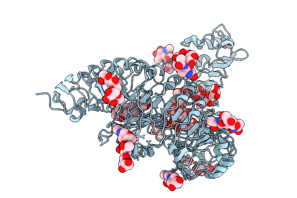
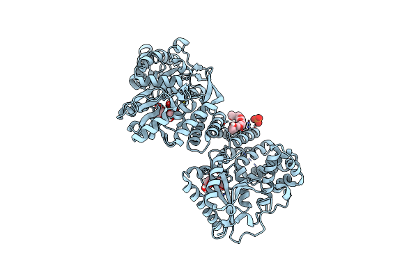
Crystal Structure Of The N-Terminal Kinase Domain From Saccharomyces Cerevisiae Vip1 In Complex With Adp.
Organism: Saccharomyces cerevisiae
Method: X-RAY DIFFRACTION Resolution:1.18 Å Release Date: 2025-02-26 Classification: BIOSYNTHETIC PROTEIN Ligands: ADP, EDO |
 |
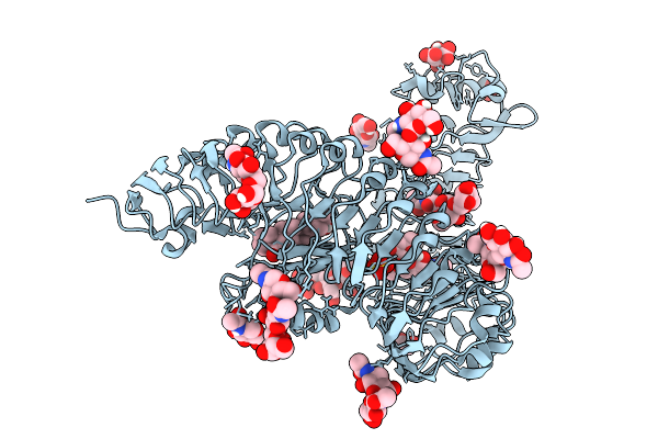
Crystal Structure Of The C-Terminal Phosphatase Domain From Saccharomyces Cerevisiae Vip1 (Apo)
Organism: Saccharomyces cerevisiae
Method: X-RAY DIFFRACTION Resolution:3.20 Å Release Date: 2025-02-26 Classification: BIOSYNTHETIC PROTEIN Ligands: ZN |
 |
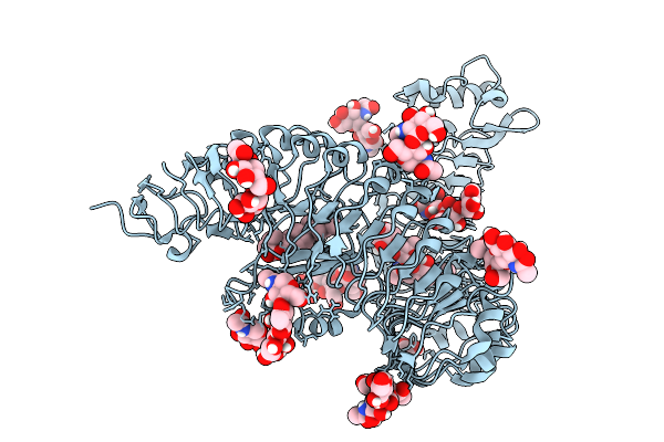
Crystal Structure Of The Engineered C-Terminal Phosphatase Domain From Saccharomyces Cerevisiae Vip1 (Apo, Loop Deletion Residues 848-918)
Organism: Saccharomyces cerevisiae
Method: X-RAY DIFFRACTION Resolution:3.40 Å Release Date: 2025-02-26 Classification: BIOSYNTHETIC PROTEIN Ligands: GOL, ZN, LPC, ACT |
 |
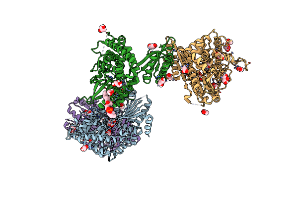
Crystal Structure Of The Engineered C-Terminal Phosphatase Domain From Saccharomyces Cerevisiae Vip1 In Complex With 1,5-Insp8 (Phosphatase Dead Mutant, Loop Deletion Residues 848-918)
Organism: Saccharomyces cerevisiae
Method: X-RAY DIFFRACTION Resolution:2.36 Å Release Date: 2025-02-26 Classification: BIOSYNTHETIC PROTEIN Ligands: ZN, SPM, ORN, PUT, EDO, I8P, 1JW |
 |
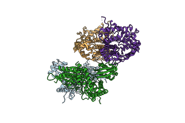
Structure Of The Fast1-Fast2-Rap Module From Human Fastkd4 By Carrier-Driven Crystallisation With Maltose Binding Protein From E. Coli.
Organism: Homo sapiens
Method: X-RAY DIFFRACTION Resolution:2.15 Å Release Date: 2025-01-15 Classification: RNA BINDING PROTEIN Ligands: P33, PO4, GLC, FRU |
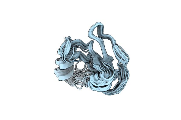 |
Nmr Solution Structure Of Thyropin Irthy-Cd From The Hard Tick Ixodes Ricinus
Organism: Ixodes ricinus
Method: SOLUTION NMR Release Date: 2024-02-28 Classification: PROTEIN BINDING |
 |
Organism: Schistosoma mansoni
Method: X-RAY DIFFRACTION Resolution:1.55 Å Release Date: 2024-02-21 Classification: HYDROLASE Ligands: UA9, EDO, NA |