
Search Count: 50
 |
Organism: Homo sapiens
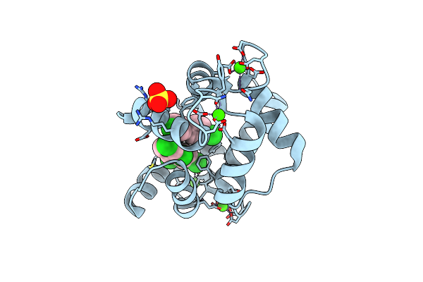
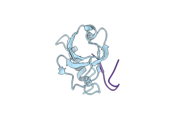
Method: X-RAY DIFFRACTION Resolution:1.90 Å Release Date: 2022-08-17 Classification: METAL BINDING PROTEIN Ligands: 85H, SO4, CA |
 |
Organism: Homo sapiens
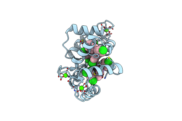
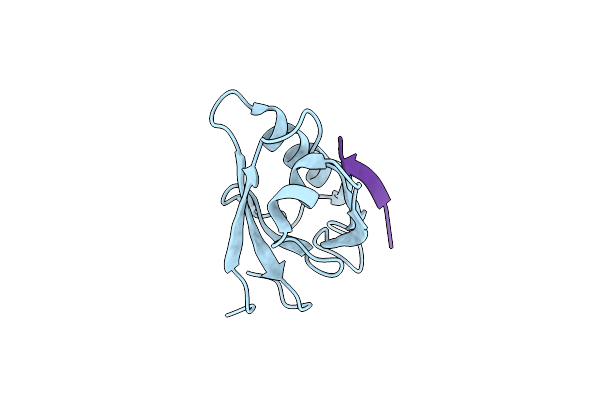
Method: X-RAY DIFFRACTION Resolution:2.28 Å Release Date: 2022-08-17 Classification: METAL BINDING PROTEIN Ligands: 85H, CA |
 |
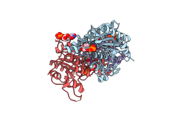
Crystal Structure Of Umpk From M. Tuberculosis In Complex With Udp And Utp (C2 Form)
Organism: Mycobacterium tuberculosis h37rv
Method: X-RAY DIFFRACTION Resolution:3.12 Å Release Date: 2022-03-02 Classification: TRANSFERASE Ligands: UTP, UDP |
 |
Crystal Structure Of Umpk From M. Tuberculosis In Complex With Udp And Utp (P21212 Form)
Organism: Mycobacterium tuberculosis h37rv
Method: X-RAY DIFFRACTION Resolution:3.33 Å Release Date: 2022-03-02 Classification: TRANSFERASE Ligands: UDP, UTP |
 |
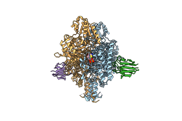
Cryoem Structure Of Mycobacterium Tuberculosis Ump Kinase (Umpk) In Complex With Udp And Utp
Organism: Mycobacterium tuberculosis
Method: ELECTRON MICROSCOPY Release Date: 2022-01-12 Classification: TRANSFERASE Ligands: UDP, UTP |
 |
Organism: Streptococcus agalactiae
Method: X-RAY DIFFRACTION Resolution:2.64 Å Release Date: 2021-08-18 Classification: SIGNALING PROTEIN Ligands: SO4, PT |
 |
Organism: Streptococcus agalactiae
Method: X-RAY DIFFRACTION Release Date: 2021-08-18 Classification: SIGNALING PROTEIN Ligands: 2BA, SO4 |
 |
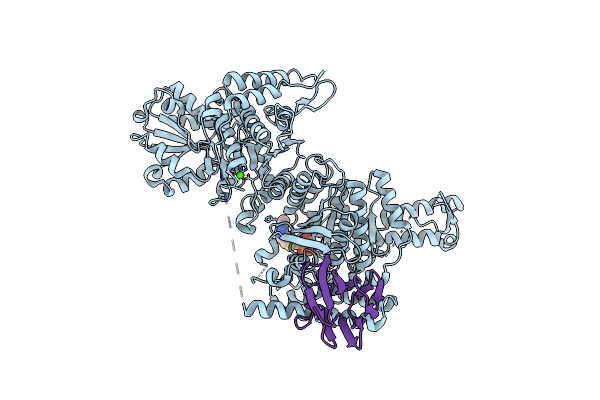
Organism: Homo sapiens, Bordetella pertussis
Method: X-RAY DIFFRACTION Resolution:3.94 Å Release Date: 2021-03-17 Classification: METAL BINDING PROTEIN Ligands: CA |
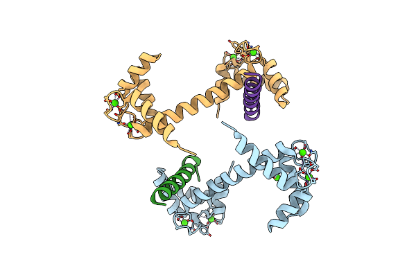 |
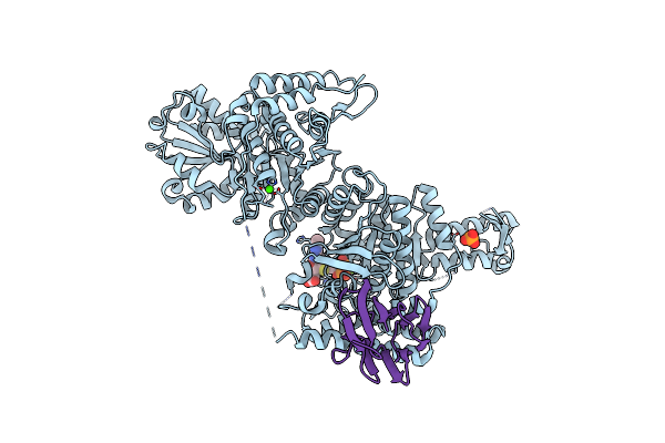
Organism: Homo sapiens, Bordetella pertussis
Method: X-RAY DIFFRACTION Resolution:3.12 Å Release Date: 2021-03-17 Classification: METAL BINDING PROTEIN Ligands: CA |
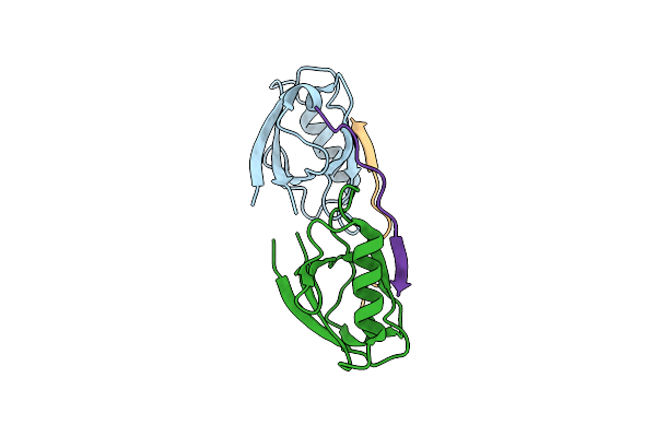 |
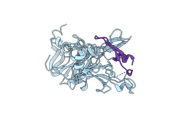
Crystal Structure Of Whirlin Pdz3 In Complex With Myosin 15A C-Terminal Pdz Binding Motif Peptide
Organism: Mus musculus
Method: X-RAY DIFFRACTION Resolution:1.70 Å Release Date: 2020-10-07 Classification: STRUCTURAL PROTEIN |
 |
Crystal Structure Of Whirlin Pdz3_C-Ter In Complex With Myosin 15A C-Terminal Pdz Binding Motif Peptide
Organism: Mus musculus
Method: X-RAY DIFFRACTION Resolution:1.93 Å Release Date: 2020-10-07 Classification: STRUCTURAL PROTEIN |
 |
Crystal Structure Of Whirlin Pdz3_C-Ter In Complex With Cask Internal Pdz Binding Motif Peptide
Organism: Mus musculus
Method: X-RAY DIFFRACTION Resolution:1.63 Å Release Date: 2020-10-07 Classification: STRUCTURAL PROTEIN |
 |
Crystal Structure Of Whirlin Pdz3_C-Ter In Complex With Harmonin A1 C-Terminal Pdz Binding Motif Peptide
Organism: Mus musculus, Rattus norvegicus
Method: X-RAY DIFFRACTION Resolution:3.17 Å Release Date: 2020-10-07 Classification: STRUCTURAL PROTEIN |
 |
Crystal Structure Of Whirlin Pdz3_C-Ter In Complex With Taperin Internal Pdz Binding Motif Peptide
Organism: Mus musculus, Homo sapiens
Method: X-RAY DIFFRACTION Resolution:1.32 Å Release Date: 2020-10-07 Classification: STRUCTURAL PROTEIN |
 |
Crystal Structure Of The Mycobacterium Tuberculosis Pknb Kinase Domain (L33E Mutant) In Complex With Its Substrate Gara
Organism: Mycobacterium tuberculosis (strain atcc 25618 / h37rv)
Method: X-RAY DIFFRACTION Resolution:2.37 Å Release Date: 2019-05-22 Classification: SIGNALING PROTEIN Ligands: ACP, MG, SO4 |
 |
Crystal Structure Of The Wild-Type Suca Domain Of Mycobacterium Smegmatis Kgd (Alpha-Ketoglutarate Decarboxylase), In Complex With Gara
Organism: Mycobacterium smegmatis (strain atcc 700084 / mc(2)155)
Method: X-RAY DIFFRACTION Resolution:2.15 Å Release Date: 2019-05-22 Classification: OXIDOREDUCTASE Ligands: TPP, MG, CA |
 |
Crystal Structure Of The Suca Domain Of Mycobacterium Smegmatis Kgd (Alpha-Ketoglutarate Decarboxylase), Mutant R802A, In Complex With Gara
Organism: Mycobacterium smegmatis (strain atcc 700084 / mc(2)155)
Method: X-RAY DIFFRACTION Resolution:2.20 Å Release Date: 2019-05-22 Classification: OXIDOREDUCTASE Ligands: TPP, MG, CA |
 |
Crystal Structure Of The Suca Domain Of Mycobacterium Smegmatis Kgd (R802A) In Complex With Gara, Following 2-Oxoglutarate Soak
Organism: Mycobacterium smegmatis (strain atcc 700084 / mc(2)155)
Method: X-RAY DIFFRACTION Resolution:2.40 Å Release Date: 2019-05-22 Classification: OXIDOREDUCTASE Ligands: MG, CA, PO4, TD6 |
 |
Crystal Structure Of Plasmodium Falciparum Ama1 In Complex With A 39 Aa Pvron2 Peptide
Organism: Plasmodium falciparum vietnam oak-knoll (fvo), Plasmodium vivax sal-1
Method: X-RAY DIFFRACTION Resolution:1.90 Å Release Date: 2017-09-06 Classification: CELL INVASION |
 |
Crystal Structure Of Plasmodium Vivax Ama1 In Complex With A 39 Aa Pvron2 Peptide
Organism: Plasmodium vivax sal-1, Plasmodium vivax
Method: X-RAY DIFFRACTION Resolution:2.15 Å Release Date: 2017-09-06 Classification: CELL INVASION |