
Search Count: 21
 |
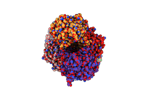
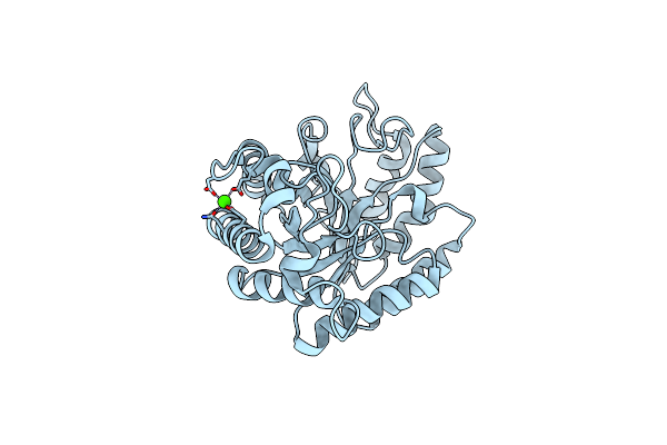
Crystal Structure Of The Gh51 Arabinofuranosidase From Xanthomonas Axonopodis Pv. Citri
Organism: Xanthomonas axonopodis pv. citri (strain 306)
Method: X-RAY DIFFRACTION Resolution:1.91 Å Release Date: 2019-02-20 Classification: HYDROLASE Ligands: GOL |
 |
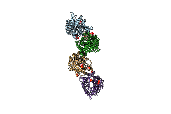
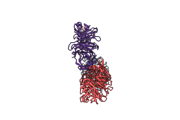
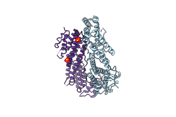
Crystal Structure Of The Gh2 Exo-Beta-Mannanase From Xanthomonas Axonopodis Pv. Citri
Organism: Xanthomonas axonopodis pv. citri (strain 306)
Method: X-RAY DIFFRACTION Resolution:1.90 Å Release Date: 2018-07-18 Classification: HYDROLASE/CARBOHYDRATE Ligands: GOL, PEG, ACT |
 |
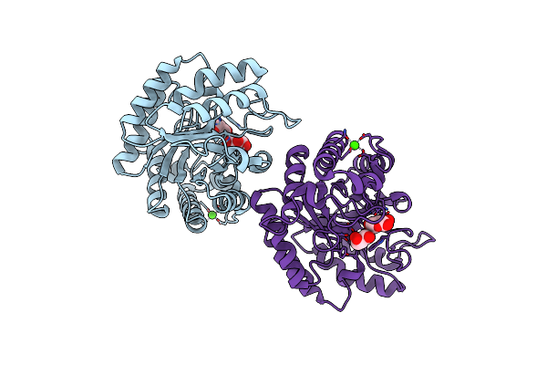
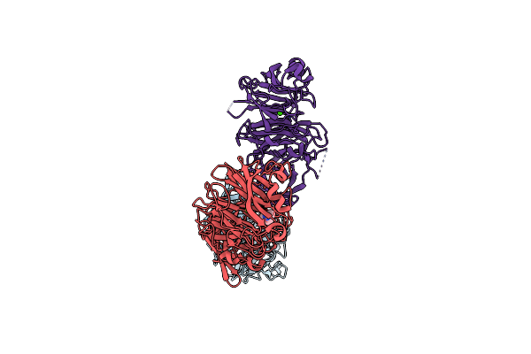
Crystal Structure Of The Gh2 Exo-Beta-Mannanase From Xanthomonas Axonopodis Pv. Citri In Complex With Mannose
Organism: Xanthomonas axonopodis pv. citri (strain 306)
Method: X-RAY DIFFRACTION Resolution:2.13 Å Release Date: 2018-07-18 Classification: HYDROLASE/CARBOHYDRATE Ligands: BMA, ACT |
 |
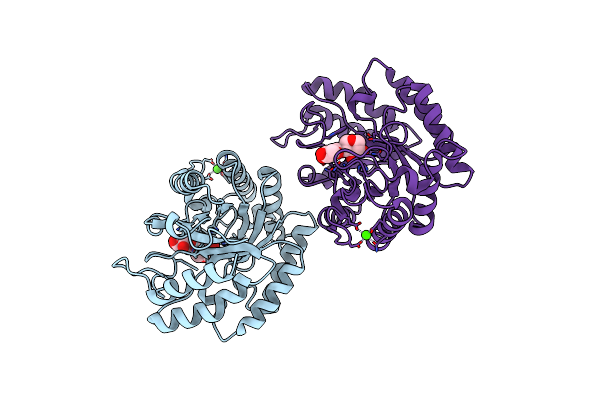
Crystal Structure Of The Nucleophile Mutant (E575A) Of The Gh2 Exo-Beta-Mannanase From Xanthomonas Axonopodis Pv. Citri
Organism: Xanthomonas axonopodis pv. citri (strain 306)
Method: X-RAY DIFFRACTION Resolution:2.00 Å Release Date: 2018-07-18 Classification: HYDROLASE/CARBOHYDRATE Ligands: BMA |
 |
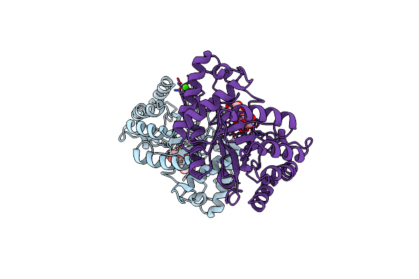
Crystal Structure Of The Acid-Base Mutant (E477A) Of The Gh2 Exo-Beta-Mannanase From Xanthomonas Axonopodis Pv. Citri
Organism: Xanthomonas axonopodis pv. citri (strain 306)
Method: X-RAY DIFFRACTION Resolution:2.20 Å Release Date: 2018-07-18 Classification: HYDROLASE/CARBOHYDRATE Ligands: BMA |
 |
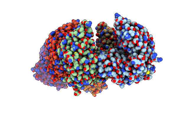
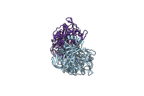
Crystal Structure Of The Gh1 Beta-Glucosidase From Exiguobacterium Antarcticum B7 In Space Group P21
Organism: Exiguobacterium antarcticum (strain b7)
Method: X-RAY DIFFRACTION Resolution:2.24 Å Release Date: 2016-04-13 Classification: HYDROLASE Ligands: SO4 |
 |
Crystal Structure Of The Gh1 Beta-Glucosidase From Exiguobacterium Antarcticum B7 In Space Group C2221
Organism: Exiguobacterium antarcticum (strain b7)
Method: X-RAY DIFFRACTION Resolution:2.15 Å Release Date: 2016-04-13 Classification: HYDROLASE Ligands: CXS, SO4, GOL |
 |
Crystal Structure Of A Novel Reducing-End Xylose-Releasing Exo-Oligoxylanase (Xyna) Belonging To Gh10 Family (Space Group P1211)
Organism: Xanthomonas axonopodis pv. citri
Method: X-RAY DIFFRACTION Resolution:2.86 Å Release Date: 2014-10-08 Classification: HYDROLASE |
 |
Crystal Structure Of A Novel Reducing-End Xylose-Releasing Exo-Oligoxylanase (Xyna) Belonging To Gh10 Family (Space Group P43212)
Organism: Xanthomonas axonopodis pv. citri
Method: X-RAY DIFFRACTION Resolution:3.00 Å Release Date: 2014-10-08 Classification: HYDROLASE |
 |
Crystal Structure Of Gh10 Endo-B-1,4-Xylanase (Xynb) From Xanthomonas Axonopodis Pv Citri In The Native Form
Organism: Xanthomonas axonopodis pv. citri
Method: X-RAY DIFFRACTION Resolution:1.30 Å Release Date: 2014-10-08 Classification: HYDROLASE Ligands: CA |
 |
Crystal Structure Of Gh10 Endo-B-1,4-Xylanase (Xynb) From Xanthomonas Axonopodis Pv Citri Complexed With Xylose
Organism: Xanthomonas axonopodis pv. citri
Method: X-RAY DIFFRACTION Resolution:1.60 Å Release Date: 2014-10-08 Classification: HYDROLASE Ligands: XYP, GOL, CA |
 |
Crystal Structure Of Gh10 Endo-B-1,4-Xylanase (Xynb) From Xanthomonas Axonopodis Pv Citri Complexed With Xylobiose
Organism: Xanthomonas axonopodis pv. citri
Method: X-RAY DIFFRACTION Resolution:1.40 Å Release Date: 2014-10-08 Classification: HYDROLASE Ligands: CA |
 |
Crystal Structure Of Gh10 Endo-B-1,4-Xylanase (Xynb) From Xanthomonas Axonopodis Pv Citri Complexed With Xylotriose
Organism: Xanthomonas axonopodis pv. citri
Method: X-RAY DIFFRACTION Resolution:1.42 Å Release Date: 2014-10-08 Classification: HYDROLASE Ligands: CA, XYP |
 |
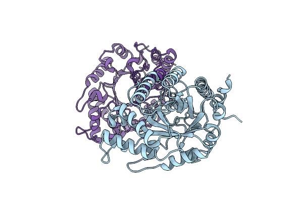
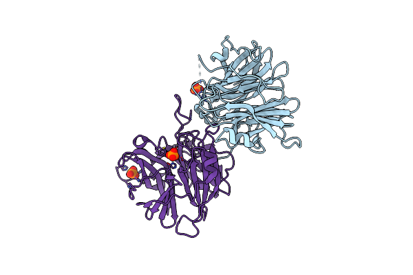
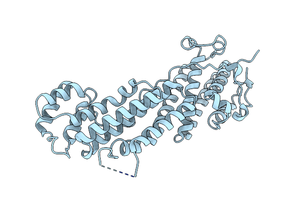
Crystal Structure Of Endo-1,5-Alpha-L-Arabinanase From Thermotoga Petrophila Rku-1
Organism: Thermotoga petrophila
Method: X-RAY DIFFRACTION Resolution:1.75 Å Release Date: 2014-02-05 Classification: HYDROLASE Ligands: PEG, CA |
 |
Crystal Structure Of Endo-1,5-Alpha-L-Arabinanase From Thermotoga Petrophila Rku-1 In Complex With Tris
Organism: Thermotoga petrophila
Method: X-RAY DIFFRACTION Resolution:1.76 Å Release Date: 2014-02-05 Classification: HYDROLASE Ligands: TRS, CA |
 |
Crystal Structure Of Endo-1,5-Alpha-L-Arabinanase From A Bovine Ruminal Metagenomic Library
Organism: Bos taurus
Method: X-RAY DIFFRACTION Resolution:1.90 Å Release Date: 2014-02-05 Classification: HYDROLASE Ligands: IOD, GOL, NA |
 |
Crystal Structure Of Exo-1,5-Alpha-L-Arabinanase From Bovine Ruminal Metagenomic Library
Organism: Uncultured bacterium
Method: X-RAY DIFFRACTION Resolution:2.90 Å Release Date: 2014-02-05 Classification: HYDROLASE Ligands: PO4 |
 |
Organism: Homo sapiens
Method: X-RAY DIFFRACTION Resolution:2.20 Å Release Date: 2013-10-09 Classification: PROTEIN TRANSPORT Ligands: SO4 |
 |
Organism: Homo sapiens
Method: X-RAY DIFFRACTION Resolution:2.07 Å Release Date: 2013-10-09 Classification: PROTEIN TRANSPORT Ligands: NO3 |
 |
Organism: Homo sapiens
Method: X-RAY DIFFRACTION Resolution:2.95 Å Release Date: 2013-10-09 Classification: PROTEIN TRANSPORT |