
Search Count: 7
 |
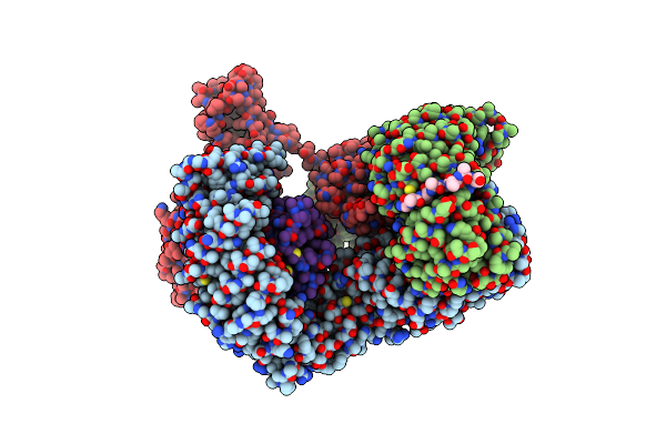
Organism: Mus musculus
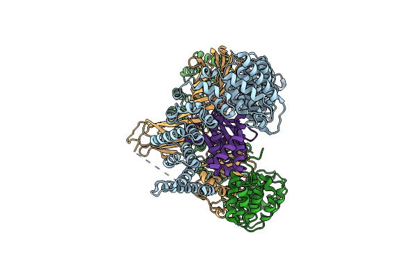
Method: ELECTRON MICROSCOPY Release Date: 2022-03-30 Classification: ENDOCYTOSIS |
 |
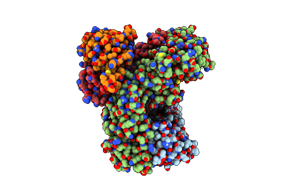
Organism: Mus musculus
Method: ELECTRON MICROSCOPY Release Date: 2022-03-30 Classification: ENDOCYTOSIS |
 |
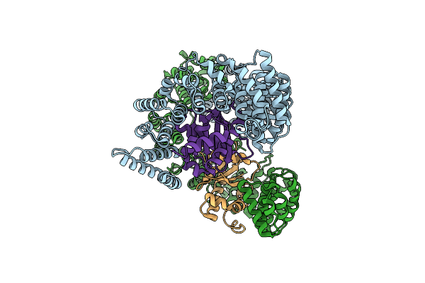
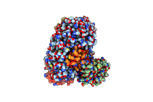
Organism: Mus musculus
Method: ELECTRON MICROSCOPY Release Date: 2022-03-30 Classification: ENDOCYTOSIS |
 |
Organism: Mus musculus
Method: ELECTRON MICROSCOPY Release Date: 2022-03-30 Classification: ENDOCYTOSIS |
 |
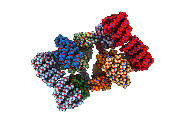
Ap2 Bound To The Apa Domain Of Sgip And Heparin; Partial Signal Subtraction And Symmetry Expansion
Organism: Mus musculus
Method: ELECTRON MICROSCOPY Release Date: 2022-03-30 Classification: ENDOCYTOSIS |
 |
Organism: Mus musculus, Rattus norvegicus
Method: ELECTRON MICROSCOPY Resolution:3.20 Å Release Date: 2019-09-11 Classification: ENDOCYTOSIS |
 |
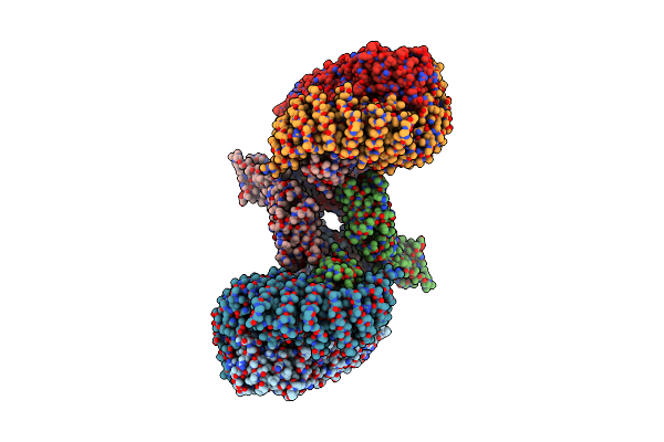
Cryo-Em Structure Of Phosphorylated Ap-2 (Mu E302K) Bound To Necap In The Presence Of Ss Dna
Organism: Mus musculus, Rattus norvegicus
Method: ELECTRON MICROSCOPY Release Date: 2019-09-11 Classification: ENDOCYTOSIS |