
Search Count: 18
 |
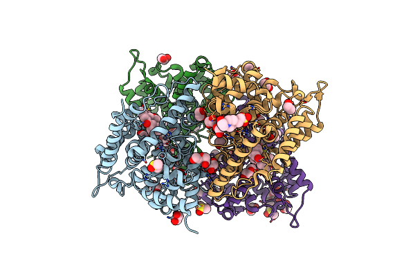
Organism: Homo sapiens
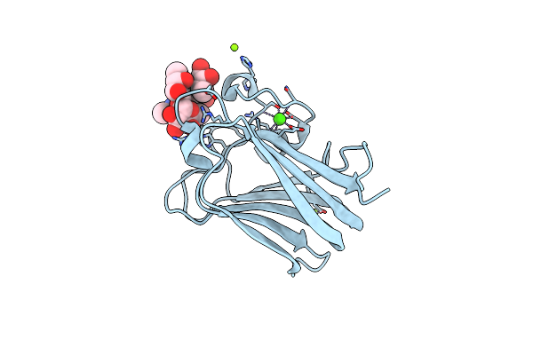
Method: X-RAY DIFFRACTION Resolution:1.52 Å Release Date: 2022-08-10 Classification: PROTEIN TRANSPORT Ligands: FQ7 |
 |
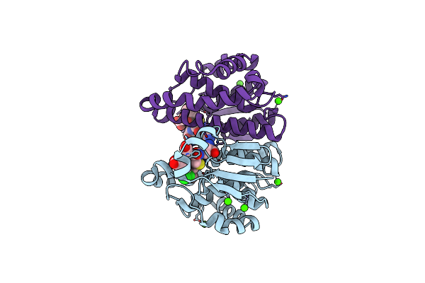
Organism: Homo sapiens
Method: X-RAY DIFFRACTION Resolution:2.03 Å Release Date: 2015-01-21 Classification: HYDROLASE/HYDROLASE INHIBITOR Ligands: 2SR, EDO, PEG, ZN, DMS, EPE |
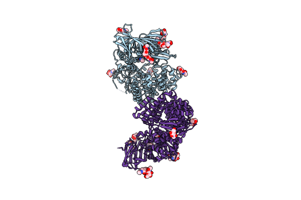 |
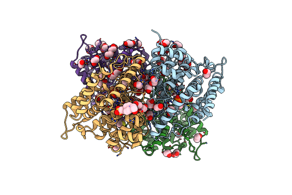
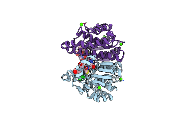
Crystal Structure Of Human Insulin Regulated Aminopeptidase With Alanine In Active Site
Organism: Homo sapiens
Method: X-RAY DIFFRACTION Resolution:3.02 Å Release Date: 2014-12-03 Classification: HYDROLASE Ligands: ZN, NAG, UNL |
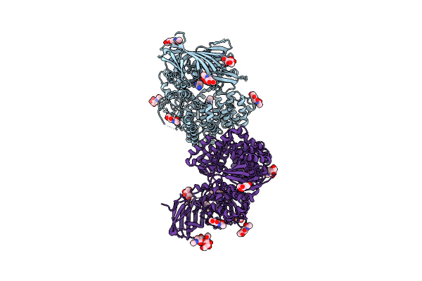 |
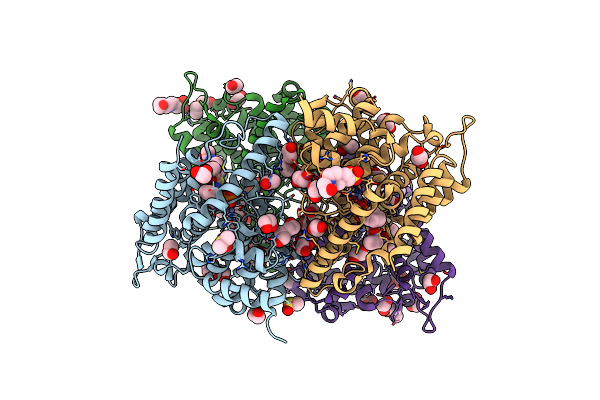
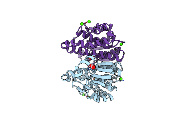
Crystal Structure Of Human Insulin Regulated Aminopeptidase With Lysine In Active Site
Organism: Homo sapiens
Method: X-RAY DIFFRACTION Resolution:2.96 Å Release Date: 2014-12-03 Classification: HYDROLASE Ligands: ZN, LYS, NAG |
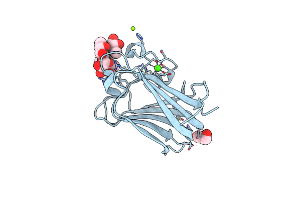 |
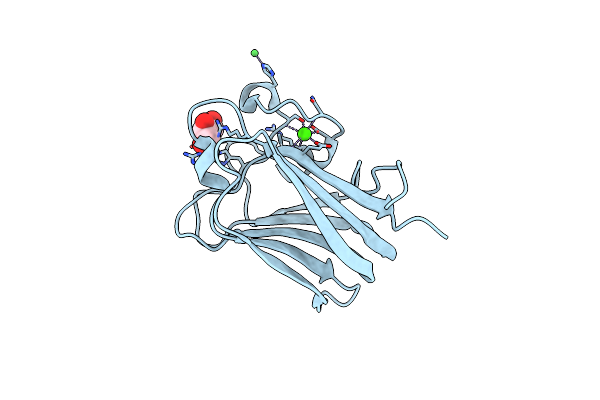
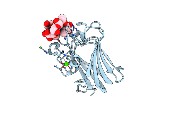
His 62 Mutant Of The Lectin Binding Domain Of Lectinolysin Complexed With Lewis Y
Organism: Streptococcus mitis
Method: X-RAY DIFFRACTION Resolution:1.60 Å Release Date: 2012-11-21 Classification: SUGAR BINDING PROTEIN Ligands: MG, GOL, CA |
 |
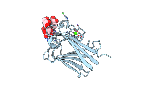
His 62 Mutant Of The Lectin Binding Domain Of Lectinolysin Complexed With Lewis B
Organism: Streptococcus mitis
Method: X-RAY DIFFRACTION Resolution:1.60 Å Release Date: 2012-11-21 Classification: SUGAR BINDING PROTEIN Ligands: MG, CA |
 |
Organism: Homo sapiens
Method: X-RAY DIFFRACTION Resolution:2.10 Å Release Date: 2011-10-26 Classification: HYDROLASE Ligands: EDO, PEG, ZN, PO4, DMS, EPE |
 |
Organism: Homo sapiens
Method: X-RAY DIFFRACTION Resolution:1.90 Å Release Date: 2011-10-26 Classification: Hydrolase/Inhibitor Ligands: ZN, EDO, DMS, PEG, PO4, JN4, EPE |
 |
Crystal Structure Of The Catalytic Domain Of Pde4D2 Complexed With Compound 10D
Organism: Homo sapiens
Method: X-RAY DIFFRACTION Resolution:2.65 Å Release Date: 2011-10-26 Classification: Hydrolase/Hydrolase Inhibitor Ligands: ZN, J25, EDO, DMS, EPE |
 |
Organism: Homo sapiens
Method: X-RAY DIFFRACTION Resolution:2.44 Å Release Date: 2011-10-26 Classification: Hydrolase/Hydrolase Inhibitor Ligands: ZN, EDO, JN8, DMS, EPE |
 |
Organism: Homo sapiens
Method: X-RAY DIFFRACTION Resolution:2.60 Å Release Date: 2011-10-26 Classification: Hydrolase/Hydrolase Inhibitor Ligands: EDO, ZN, PEG, JN7, PO4, DMS |
 |
Organism: Streptococcus mitis
Method: X-RAY DIFFRACTION Resolution:1.91 Å Release Date: 2010-12-29 Classification: BLOOD CLOTTING Ligands: CA, NI, GOL |
 |
Organism: Streptococcus mitis
Method: X-RAY DIFFRACTION Resolution:2.01 Å Release Date: 2010-12-29 Classification: BLOOD CLOTTING Ligands: CA, NI |
 |
Organism: Streptococcus mitis
Method: X-RAY DIFFRACTION Resolution:1.90 Å Release Date: 2010-12-29 Classification: BLOOD CLOTTING Ligands: FUC, CA, NI |
 |
Organism: Streptococcus mitis
Method: X-RAY DIFFRACTION Resolution:2.00 Å Release Date: 2010-12-29 Classification: BLOOD CLOTTING Ligands: CA, NI |
 |
Crystal Structure Of The Human Gst Pi C47S/Y108V Double Mutant In Complex With The Ethacrynic Acid-Glutathione Conjugate
Organism: Homo sapiens
Method: X-RAY DIFFRACTION Resolution:2.10 Å Release Date: 2010-03-23 Classification: TRANSFERASE Ligands: EAA, GSH, CA |
 |
Organism: Homo sapiens
Method: X-RAY DIFFRACTION Resolution:1.80 Å Release Date: 2010-03-23 Classification: TRANSFERASE Ligands: CA, CO3, PO4 |
 |
Crystal Structure Of The Human Gst Pi C47S/Y108V Double Mutant In Complex With The Ethacrynic Acid-Glutathione Conjugate (Grown In The Absence Of The Reducing Agent Dtt)
Organism: Homo sapiens
Method: X-RAY DIFFRACTION Resolution:2.60 Å Release Date: 2010-03-23 Classification: TRANSFERASE Ligands: GSH, CA, EAA |