
Search Count: 7
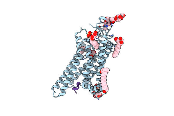 |
Crystal Structure Of The Active G-Protein-Coupled Receptor Opsin In Complex With The Finger-Loop Peptide Derived From The Full-Length Arrestin-1
Organism: Bos taurus
Method: X-RAY DIFFRACTION Resolution:2.75 Å Release Date: 2014-09-17 Classification: SIGNALING PROTEIN Ligands: BOG, PLM, SO4, ACT |
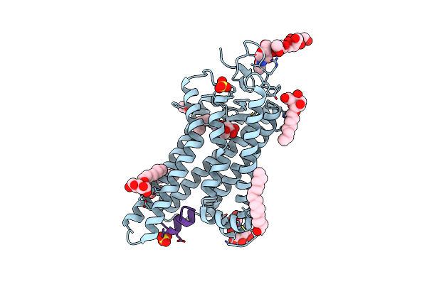 |
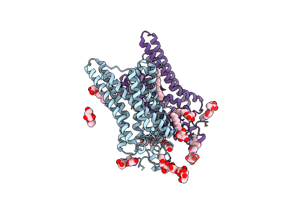
Crystal Structure Of Active Conformation Of Gpcr Opsin Stabilized By Octylglucoside
Organism: Bos taurus
Method: X-RAY DIFFRACTION Resolution:2.65 Å Release Date: 2013-10-30 Classification: SIGNALING PROTEIN Ligands: BOG, PLM, SO4, ACT |
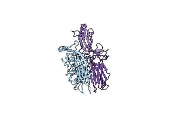 |
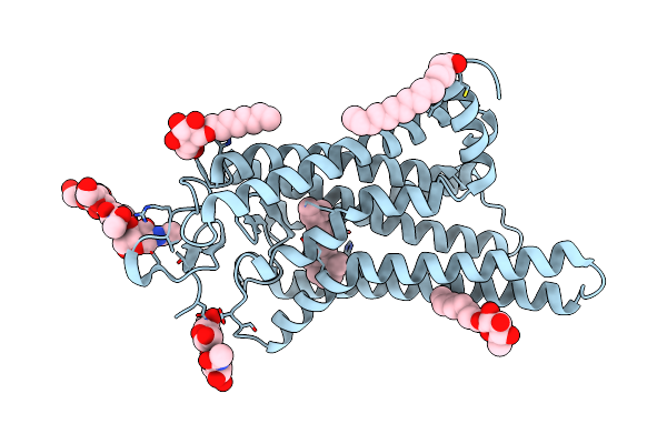
Crystal Structure Of C-Terminally Truncated Arrestin Reveals Mechanism Of Arrestin Activation
Organism: Bos taurus
Method: X-RAY DIFFRACTION Resolution:3.00 Å Release Date: 2013-05-01 Classification: SIGNALING PROTEIN |
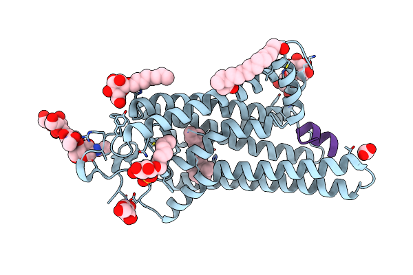 |
Crystal Structure Of Metarhodopsin Ii In Complex With A C-Terminal Peptide Derived From The Galpha Subunit Of Transducin
Organism: Bos taurus
Method: X-RAY DIFFRACTION Resolution:2.85 Å Release Date: 2011-03-09 Classification: SIGNALING PROTEIN Ligands: RET, NAG, BOG, PLM, SO4, ACT |
 |
Organism: Bos taurus
Method: X-RAY DIFFRACTION Resolution:3.00 Å Release Date: 2011-03-09 Classification: SIGNALING PROTEIN Ligands: BOG, PLM, RET |
 |
Crystal Structure Of The Active G-Protein-Coupled Receptor Opsin In Complex With A C-Terminal Peptide Derived From The Galpha Subunit Of Transducin
Organism: Bos taurus
Method: X-RAY DIFFRACTION Resolution:3.20 Å Release Date: 2008-09-23 Classification: SIGNALING PROTEIN Ligands: BOG, PLM |
 |
Crystal Structure Of Native Opsin: The G Protein-Coupled Receptor Rhodopsin In Its Ligand-Free State
Organism: Bos taurus
Method: X-RAY DIFFRACTION Resolution:2.90 Å Release Date: 2008-06-24 Classification: SIGNALING PROTEIN Ligands: BGL, PLM |