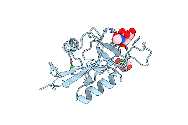
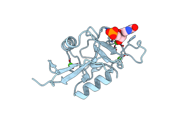
Search Count: 80
 |
Organism: Drosophila melanogaster, Homo sapiens
Method: ELECTRON MICROSCOPY Release Date: 2025-10-29 Classification: LIGASE Ligands: AMP |
 |
Organism: Drosophila melanogaster, Homo sapiens
Method: ELECTRON MICROSCOPY Release Date: 2025-10-29 Classification: LIGASE Ligands: AMP |
 |
Organism: Drosophila melanogaster, Homo sapiens
Method: ELECTRON MICROSCOPY Release Date: 2025-10-29 Classification: LIGASE Ligands: AMP |
 |
Consensus Structure Of Uba6-Ubdha-Birc6 Trapped Ternary Complex (Singly Loaded)
Organism: Homo sapiens
Method: ELECTRON MICROSCOPY Release Date: 2025-10-29 Classification: LIGASE Ligands: IHP, ATP |
 |
Consensus Structure Of Uba6-Ubdha-Birc6 Trapped Ternary Complex (Doubly Loaded)
Organism: Homo sapiens
Method: ELECTRON MICROSCOPY Release Date: 2025-10-29 Classification: LIGASE Ligands: IHP, AMP |
 |
Organism: Homo sapiens
Method: ELECTRON MICROSCOPY Release Date: 2025-10-29 Classification: LIGASE Ligands: IHP, AMP |
 |
Organism: Homo sapiens
Method: ELECTRON MICROSCOPY Release Date: 2025-10-29 Classification: LIGASE Ligands: IHP, AMP |
 |
Organism: Homo sapiens
Method: ELECTRON MICROSCOPY Release Date: 2025-10-29 Classification: LIGASE Ligands: IHP, ATP |
 |
Organism: Homo sapiens
Method: ELECTRON MICROSCOPY Release Date: 2025-10-29 Classification: LIGASE Ligands: IHP, ATP |
 |
Organism: Homo sapiens
Method: ELECTRON MICROSCOPY Release Date: 2025-10-29 Classification: LIGASE Ligands: IHP, ATP |
 |
Organism: Homo sapiens
Method: ELECTRON MICROSCOPY Release Date: 2025-10-29 Classification: LIGASE Ligands: IHP, ATP |
 |
Organism: Homo sapiens
Method: ELECTRON MICROSCOPY Release Date: 2025-10-29 Classification: LIGASE Ligands: IHP, ATP |
 |
Organism: Homo sapiens
Method: ELECTRON MICROSCOPY Release Date: 2025-10-29 Classification: LIGASE Ligands: IHP, ATP |
 |
Organism: Homo sapiens
Method: ELECTRON MICROSCOPY Release Date: 2025-10-29 Classification: LIGASE Ligands: IHP, ATP |
 |
Organism: Burkholderia pyrrocinia, Homo sapiens
Method: X-RAY DIFFRACTION Resolution:2.56 Å Release Date: 2025-04-23 Classification: HYDROLASE |
 |
Organism: Parachlamydia sp., Homo sapiens
Method: X-RAY DIFFRACTION Resolution:2.15 Å Release Date: 2025-04-23 Classification: HYDROLASE Ligands: TLA |
 |
Organism: Homo sapiens, Parachlamydia sp.
Method: X-RAY DIFFRACTION Resolution:2.18 Å Release Date: 2025-04-23 Classification: HYDROLASE Ligands: CIT |
 |
Organism: Pigmentiphaga aceris, Homo sapiens
Method: X-RAY DIFFRACTION Resolution:1.89 Å Release Date: 2025-04-23 Classification: HYDROLASE Ligands: AYE |
 |
Organism: Homo sapiens
Method: X-RAY DIFFRACTION Resolution:1.20 Å Release Date: 2025-03-19 Classification: SUGAR BINDING PROTEIN Ligands: CA, GOL, A2G, NGA |
 |
Organism: Homo sapiens
Method: X-RAY DIFFRACTION Resolution:1.59 Å Release Date: 2025-03-19 Classification: SUGAR BINDING PROTEIN Ligands: CA, IMP |

