
Search Count: 48
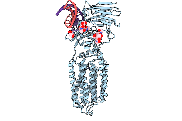 |
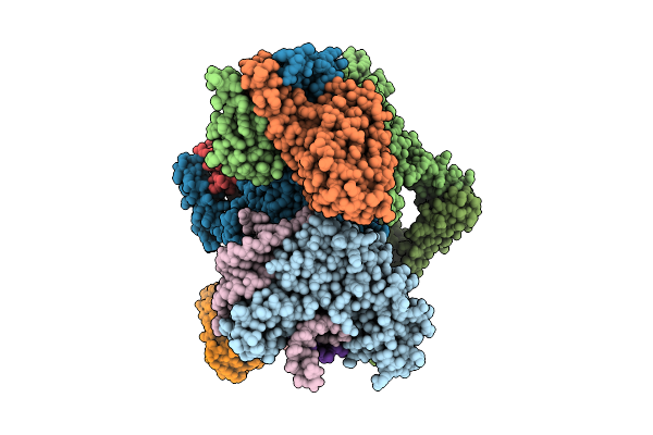
Organism: Caenorhabditis elegans, Synthetic construct
Method: ELECTRON MICROSCOPY Resolution:3.99 Å Release Date: 2026-01-28 Classification: MEMBRANE PROTEIN/RNA Ligands: NAG, ZN |
 |
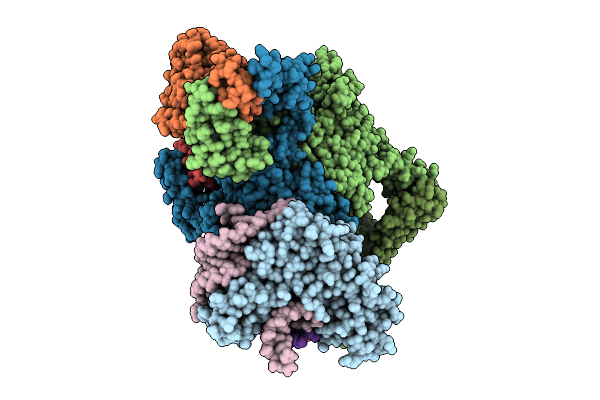
Organism: Caenorhabditis elegans, Synthetic construct
Method: ELECTRON MICROSCOPY Release Date: 2026-01-21 Classification: MEMBRANE PROTEIN/RNA Ligands: NAG, ZN, Y01, LBN |
 |
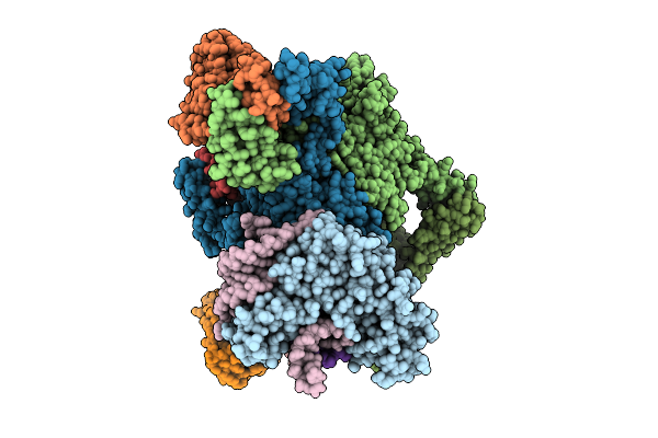
Organism: Caenorhabditis elegans
Method: ELECTRON MICROSCOPY Release Date: 2026-01-21 Classification: MEMBRANE PROTEIN Ligands: NAG, ZN, Y01, LBN |
 |
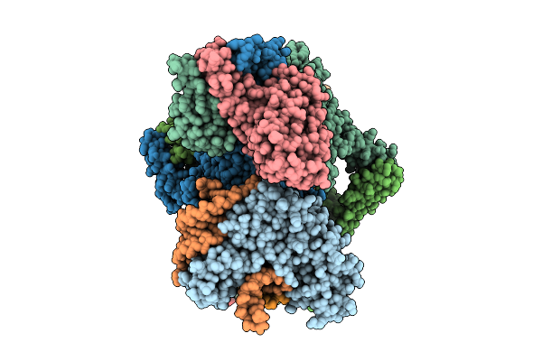
Organism: Homo sapiens
Method: ELECTRON MICROSCOPY Resolution:2.70 Å Release Date: 2025-12-10 Classification: TRANSPORT PROTEIN Ligands: A1L6U, NAG |
 |
Organism: Homo sapiens
Method: ELECTRON MICROSCOPY Resolution:2.87 Å Release Date: 2025-12-10 Classification: TRANSPORT PROTEIN Ligands: NAG, A1L6V |
 |
Organism: Homo sapiens
Method: ELECTRON MICROSCOPY Resolution:2.71 Å Release Date: 2025-12-10 Classification: TRANSPORT PROTEIN Ligands: NAG, A1L6W |
 |
Organism: Homo sapiens
Method: ELECTRON MICROSCOPY Resolution:2.66 Å Release Date: 2025-12-10 Classification: TRANSPORT PROTEIN Ligands: NAG, NA |
 |
Organism: Homo sapiens
Method: ELECTRON MICROSCOPY Resolution:2.77 Å Release Date: 2025-12-10 Classification: TRANSPORT PROTEIN |
 |
Organism: Homo sapiens
Method: ELECTRON MICROSCOPY Resolution:2.82 Å Release Date: 2025-12-10 Classification: TRANSPORT PROTEIN Ligands: A1L6U |
 |
Organism: Homo sapiens
Method: ELECTRON MICROSCOPY Resolution:2.80 Å Release Date: 2025-12-10 Classification: TRANSPORT PROTEIN Ligands: NAG, A1L6V |
 |
Organism: Homo sapiens
Method: ELECTRON MICROSCOPY Resolution:2.68 Å Release Date: 2025-12-10 Classification: TRANSPORT PROTEIN Ligands: NAG, A1L6W |
 |
Organism: Homo sapiens
Method: ELECTRON MICROSCOPY Resolution:2.80 Å Release Date: 2025-12-10 Classification: TRANSPORT PROTEIN Ligands: NAG |
 |
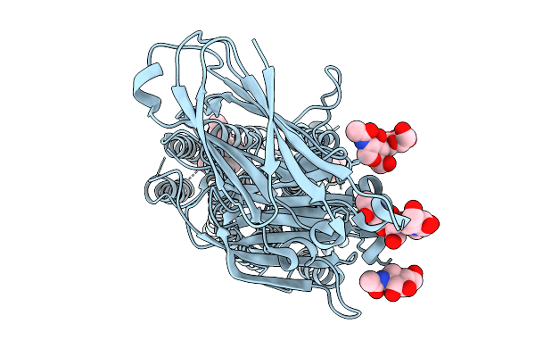
Organism: Escherichia coli
Method: ELECTRON MICROSCOPY Release Date: 2025-11-12 Classification: RNA BINDING PROTEIN Ligands: ADP, ZN, MG |
 |
Organism: Escherichia coli
Method: ELECTRON MICROSCOPY Release Date: 2025-11-12 Classification: RNA BINDING PROTEIN Ligands: ADP, ZN, MG |
 |
Organism: Escherichia coli
Method: ELECTRON MICROSCOPY Release Date: 2025-11-12 Classification: RNA BINDING PROTEIN Ligands: ADP, ZN, MG |
 |
Organism: Escherichia coli
Method: ELECTRON MICROSCOPY Release Date: 2025-11-12 Classification: RNA BINDING PROTEIN Ligands: ADP, ZN, MG |
 |
Organism: Escherichia coli
Method: ELECTRON MICROSCOPY Release Date: 2025-11-12 Classification: RNA BINDING PROTEIN Ligands: ADP, ZN, MG |
 |
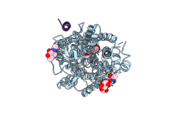
Organism: Mus musculus
Method: ELECTRON MICROSCOPY Resolution:2.70 Å Release Date: 2024-07-31 Classification: HYDROLASE Ligands: A1L0C |
 |
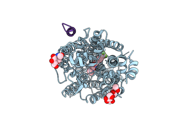
Organism: Canis lupus familiaris
Method: ELECTRON MICROSCOPY Resolution:2.20 Å Release Date: 2024-07-31 Classification: HYDROLASE Ligands: A1L0D |
 |
Organism: Canis lupus familiaris
Method: ELECTRON MICROSCOPY Resolution:2.00 Å Release Date: 2024-07-31 Classification: HYDROLASE Ligands: A1L0C |