Search Count: 23
 |
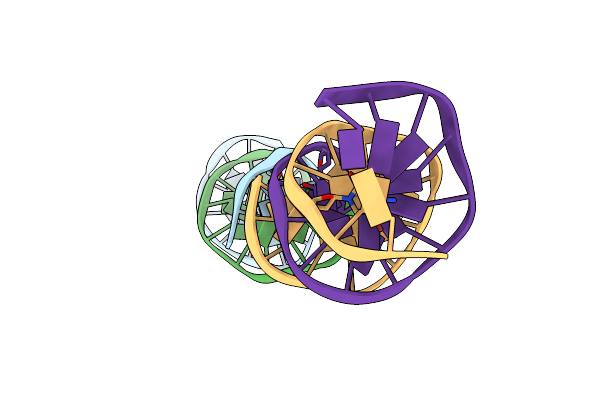
The Complex Of Cag Repeat Sequence-Specific Binding Cpip And Dsdna With A-A Mismatch
Organism: Synthetic construct
Method: X-RAY DIFFRACTION Resolution:2.80 Å Release Date: 2024-06-05 Classification: DNA Ligands: A1LYO |
 |
Organism: Homo sapiens, Mus musculus
Method: X-RAY DIFFRACTION Resolution:2.50 Å Release Date: 2022-01-05 Classification: IMMUNE SYSTEM Ligands: EDO, PEG |
 |
Organism: Human immunodeficiency virus 1
Method: X-RAY DIFFRACTION Resolution:1.00 Å Release Date: 2019-05-22 Classification: HYDROLASE/HYDROLASE INHIBITOR Ligands: GOL, SO4, B0F, CL |
 |
Organism: Human immunodeficiency virus 1
Method: X-RAY DIFFRACTION Resolution:0.85 Å Release Date: 2018-07-18 Classification: HYDROLASE/HYDROLASE INHIBITOR Ligands: 8Z0, GOL |
 |
Crystal Structure Of Kni-10343 Bound Plasmepsin Ii (Pmii) From Plasmodium Falciparum
Organism: Plasmodium falciparum
Method: X-RAY DIFFRACTION Resolution:2.00 Å Release Date: 2018-07-11 Classification: HYDROLASE Ligands: 8V9, CPS, GOL, PO4 |
 |
Crystal Structure Of Kni-10743 Bound Plasmepsin Ii (Pmii) From Plasmodium Falciparum
Organism: Plasmodium falciparum
Method: X-RAY DIFFRACTION Resolution:2.15 Å Release Date: 2018-07-11 Classification: HYDROLASE Ligands: 8VC, GOL, EDO, CPS |
 |
Crystal Structure Of Kni-10333 Bound Plasmepsin Ii (Pmii) From Plasmodium Falciparum
Organism: Plasmodium falciparum
Method: X-RAY DIFFRACTION Resolution:1.90 Å Release Date: 2018-07-11 Classification: HYDROLASE Ligands: 8VO, CPS, GOL |
 |
Crystal Structure Of Kni-10395 Bound Plasmepsin Ii (Pmii) From Plasmodium Falciparum
Organism: Plasmodium falciparum
Method: X-RAY DIFFRACTION Resolution:2.10 Å Release Date: 2018-07-11 Classification: HYDROLASE Ligands: K95, CPS, NA |
 |
Crystal Structure Of Kni-10742 Bound Plasmepsin Ii (Pmii) From Plasmodium Falciparum
Organism: Plasmodium falciparum (isolate 3d7)
Method: X-RAY DIFFRACTION Resolution:2.10 Å Release Date: 2018-07-11 Classification: HYDROLASE Ligands: CPS, 8VF, NA |
 |
Organism: Human immunodeficiency virus 1
Method: X-RAY DIFFRACTION Resolution:1.50 Å Release Date: 2018-07-11 Classification: HYDROLASE/HYDROLASE INHIBITOR Ligands: 8Z0, GOL |
 |
Organism: Homo sapiens
Method: X-RAY DIFFRACTION Resolution:1.38 Å Release Date: 2017-05-31 Classification: HYDROLASE/INHIBITOR Ligands: 89S |
 |
Organism: Homo sapiens
Method: X-RAY DIFFRACTION Resolution:1.20 Å Release Date: 2017-05-31 Classification: HYDROLASE/INHIBITOR Ligands: 89M |
 |
Crystal Structure Of Histo-Aspartic Protease (Hap) Zymogen From Plasmodium Falciparum
Organism: Plasmodium falciparum
Method: X-RAY DIFFRACTION Resolution:2.10 Å Release Date: 2011-10-12 Classification: HYDROLASE Ligands: EDO |
 |
Crystal Structure Of Kni-10395 Bound Histo-Aspartic Protease (Hap) From Plasmodium Falciparum
Organism: Plasmodium falciparum
Method: X-RAY DIFFRACTION Resolution:2.50 Å Release Date: 2011-10-12 Classification: HYDROLASE/HYDROLASE INHIBITOR Ligands: K95, EDO, PG4, ACT, PG5, NA |
 |
Organism: Plasmodium falciparum
Method: X-RAY DIFFRACTION Resolution:2.40 Å Release Date: 2011-05-11 Classification: HYDROLASE |
 |
Crystal Structure Of Kni-10006 Complex Of Plasmepsin I (Pmi) From Plasmodium Falciparum
Organism: Plasmodium falciparum
Method: X-RAY DIFFRACTION Resolution:3.10 Å Release Date: 2011-05-11 Classification: HYDROLASE/HYDROLASE INHIBITOR Ligands: 006, GOL |
 |
Crystal Structure Of Hiv-1 Protease (Q7K, L33I, L63I) In Complex With Kni-10074
Organism: Human immunodeficiency virus type 1
Method: X-RAY DIFFRACTION Resolution:2.20 Å Release Date: 2010-03-16 Classification: HYDROLASE/HYDROLASE INHIBITOR Ligands: JZP, GOL, CL |
 |
Organism: Human immunodeficiency virus 1
Method: X-RAY DIFFRACTION Resolution:0.88 Å Release Date: 2010-03-02 Classification: HYDROLASE/HYDROLASE INHIBITOR Ligands: GOL, KNJ |
 |
Crystal Structure Of Hiv-1 Protease (Q7K, L33I, L63I) In Complex With Kni-10006
Organism: Human immunodeficiency virus type 1
Method: X-RAY DIFFRACTION Resolution:1.66 Å Release Date: 2010-03-02 Classification: HYDROLASE/HYDROLASE INHIBITOR Ligands: GOL, 006 |
 |
Crystal Structure Of Hiv-1 Protease (Q7K, L33I, L63I) In Complex With Kni-10265
Organism: Human immunodeficiency virus type 1
Method: X-RAY DIFFRACTION Resolution:1.80 Å Release Date: 2010-03-02 Classification: HYDROLASE/HYDROLASE INHIBITOR Ligands: JZQ, GOL |

