
Search Count: 53
 |
Organism: Methanosarcina mazei go1, Synthetic construct
Method: X-RAY DIFFRACTION Resolution:2.11 Å Release Date: 2023-11-22 Classification: LYASE Ligands: SO4, FDA |
 |
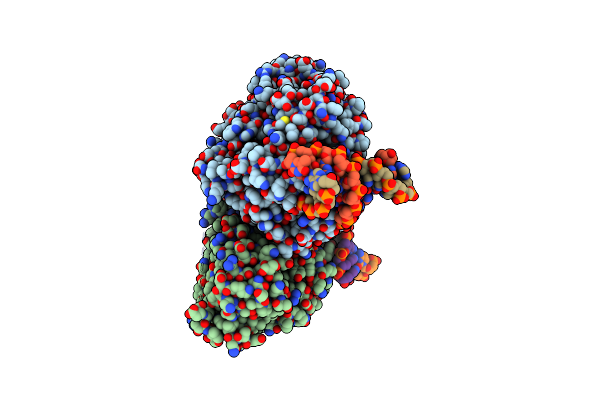
Time-Resolved Sfx Structure Of The Class Ii Photolyase Complexed With A Thymine Dimer (3 Picosecond Pump-Probe Delay)
Organism: Methanosarcina mazei go1, Synthetic construct
Method: X-RAY DIFFRACTION Resolution:2.16 Å Release Date: 2023-11-22 Classification: LYASE Ligands: SO4, FDA |
 |
Time-Resolved Sfx Structure Of The Class Ii Photolyase Complexed With A Thymine Dimer (300 Ps Pump-Probe Delay)
Organism: Methanosarcina mazei go1, Synthetic construct
Method: X-RAY DIFFRACTION Resolution:2.35 Å Release Date: 2023-11-22 Classification: LYASE Ligands: SO4, FDA |
 |
Time-Resolved Sfx Structure Of The Class Ii Photolyase Complexed With A Thymine Dimer (1 Nanosecond Pump-Probe Delay)
Organism: Methanosarcina mazei go1, Synthetic construct
Method: X-RAY DIFFRACTION Resolution:2.27 Å Release Date: 2023-11-22 Classification: LYASE Ligands: SO4, FDA |
 |
Time-Resolved Sfx Structure Of The Class Ii Photolyase Complexed With A Thymine Dimer (3 Nanosecond Pump-Probe Delay)
Organism: Methanosarcina mazei go1, Synthetic construct
Method: X-RAY DIFFRACTION Resolution:2.35 Å Release Date: 2023-11-22 Classification: LYASE Ligands: FDA, SO4 |
 |
Time-Resolved Sfx Structure Of The Class Ii Photolyase Complexed With A Thymine Dimer (10 Nanosecond Pump-Probe Delay)
Organism: Methanosarcina mazei go1, Synthetic construct
Method: X-RAY DIFFRACTION Resolution:2.36 Å Release Date: 2023-11-22 Classification: LYASE Ligands: FDA, SO4 |
 |
Time-Resolved Sfx Structure Of The Class Ii Photolyase Complexed With A Thymine Dimer (30 Nanosecond Timepoint)
Organism: Methanosarcina mazei go1, Synthetic construct
Method: X-RAY DIFFRACTION Resolution:2.39 Å Release Date: 2023-11-22 Classification: LYASE Ligands: SO4, FDA |
 |
Time-Resolved Sfx Structure Of The Class Ii Photolyase Complexed With A Thymine Dimer (1 Microsecond Pump-Probe Delay)
Organism: Methanosarcina mazei go1, Synthetic construct
Method: X-RAY DIFFRACTION Resolution:2.24 Å Release Date: 2023-11-22 Classification: LYASE Ligands: SO4, FDA |
 |
Time-Resolved Sfx Structure Of The Class Ii Photolyase Complexed With A Thymine Dimer (10 Microsecond Pump Probe Delay)
Organism: Methanosarcina mazei go1, Synthetic construct
Method: X-RAY DIFFRACTION Resolution:2.18 Å Release Date: 2023-11-22 Classification: LYASE Ligands: SO4, FDA |
 |
Time-Resolved Sfx Structure Of The Class Ii Photolyase Complexed With A Thymine Dimer (30 Microsecond Pump-Probe Delay)
Organism: Methanosarcina mazei go1, Synthetic construct
Method: X-RAY DIFFRACTION Resolution:2.25 Å Release Date: 2023-11-22 Classification: LYASE Ligands: SO4, FDA |
 |
Time-Resolved Sfx Structure Of The Class Ii Photolyase Complexed With A Thymine Dimer (100 Microsecond Timpeoint)
Organism: Methanosarcina mazei go1, Synthetic construct
Method: X-RAY DIFFRACTION Resolution:2.50 Å Release Date: 2023-11-22 Classification: LYASE Ligands: SO4, FDA |
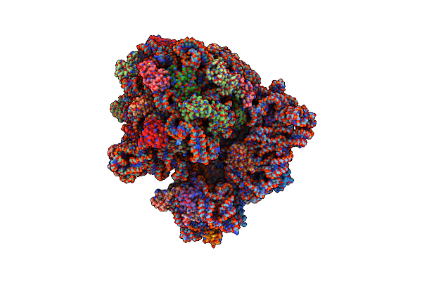 |
Cryo-Em Structure Of The E.Coli 70S Ribosome In Complex With The Antibiotic Myxovalargin B.
Organism: Myxococcus fulvus, Escherichia coli k-12
Method: ELECTRON MICROSCOPY Release Date: 2023-01-25 Classification: RIBOSOME Ligands: MG, ZN, FME, SPD |
 |
Cryo-Em Structure Of The E.Coli 50S Ribosomal Subunit In Complex With The Antibiotic Myxovalargin A.
Organism: Myxococcus fulvus, Escherichia coli k-12
Method: ELECTRON MICROSCOPY Release Date: 2023-01-18 Classification: RIBOSOME Ligands: MG, ZN |
 |
Organism: Escherichia coli, Escherichia coli k-12, Synthetic construct
Method: ELECTRON MICROSCOPY Release Date: 2023-01-11 Classification: STRUCTURAL PROTEIN |
 |
Organism: Escherichia coli, Escherichia coli k-12, Synthetic construct
Method: ELECTRON MICROSCOPY Release Date: 2023-01-11 Classification: STRUCTURAL PROTEIN |
 |
Yeast 20S Proteasome In Complex With The Covalently Bound Inhibitor B-Lactone (2R,3S)-3-Isopropyl-4-Oxo-2-Oxetane-Carboxylate (Ioc)
Organism: Saccharomyces cerevisiae
Method: X-RAY DIFFRACTION Resolution:3.00 Å Release Date: 2022-04-13 Classification: HYDROLASE Ligands: MG, CL, V08 |
 |
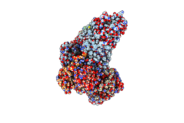
The Structure Of The Small Subunit Of The Mitoribosome From Neurospora Crassa
Organism: Neurospora crassa or74a
Method: ELECTRON MICROSCOPY Release Date: 2020-11-11 Classification: TRANSLATION Ligands: MG, ATP, K |
 |
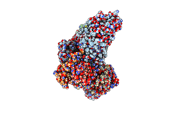
The Structure Of The Mitoribosome From Neurospora Crassa In The P/E Trna Bound State
Organism: Neurospora crassa
Method: ELECTRON MICROSCOPY Release Date: 2020-11-11 Classification: TRANSLATION Ligands: MG, SPM, K, ZN, NAD, ATP |
 |
The Structure Of The Large Subunit Of The Mitoribosome From Neurospora Crassa
Organism: Neurospora crassa or74a
Method: ELECTRON MICROSCOPY Release Date: 2020-11-11 Classification: TRANSLATION Ligands: MG, SPM, K, ZN, NAD |
 |
The Structure Of The Atp25 Bound Assembly Intermediate Of The Mitoribosome From Neurospora Crassa
Organism: Neurospora crassa or74a
Method: ELECTRON MICROSCOPY Release Date: 2020-11-11 Classification: TRANSLATION Ligands: MG, NAD, SPM, K, ZN |