
Search Count: 50
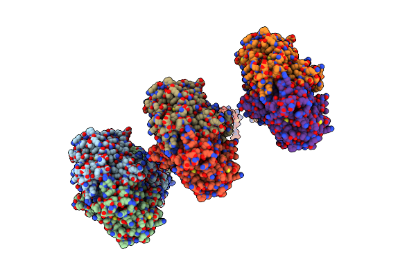 |
Organism: Homo sapiens
Method: ELECTRON MICROSCOPY Release Date: 2024-01-17 Classification: ANTIMICROBIAL PROTEIN Ligands: AF3, MG, GDP |
 |
Structure Of P. Aeruginosa Lpxc With Compound 8: (2Rs)-4-(5-(2-Fluoro-4-Methoxyphenyl)-2-Oxooxazol-3(2H)-Yl)-N-Hydroxy-2-Methyl-2-(Methylsulfonyl)Butanamide
Organism: Pseudomonas aeruginosa lesb58
Method: X-RAY DIFFRACTION Resolution:1.75 Å Release Date: 2019-12-18 Classification: HYDROLASE Ligands: ZN, H2N, GOL |
 |
Structure Of P. Aeruginosa Lpxc With Compound 10: (2Rs)-4-(5-(2-Fluoro-4-Methoxyphenyl)-1-Oxoisoindolin-2-Yl)-N-Hydroxy-2-Methyl-2-(Methylsulfonyl)Butanamide
Organism: Pseudomonas aeruginosa (strain lesb58)
Method: X-RAY DIFFRACTION Resolution:1.90 Å Release Date: 2019-12-18 Classification: HYDROLASE Ligands: ZN, UNY, UNZ |
 |
Structure Of P. Aeruginosa Lpxc With Compound 12: (2R)-4-(6-(2-Fluoro-4-Methoxyphenyl)-3-Oxo-1H-Pyrrolo[1,2-C]Imidazol-2(3H)-Yl)-N-Hydroxy-2-Methyl-2-(Methylsulfonyl)Butanamide
Organism: Pseudomonas aeruginosa lesb58
Method: X-RAY DIFFRACTION Resolution:2.20 Å Release Date: 2019-12-18 Classification: HYDROLASE Ligands: ZN, H2H, EDO |
 |
Structure Of P. Aeruginosa Lpxc With Compound 17A: (2R)-N-Hydroxy-2-Methyl-2-(Methylsulfonyl)-4(6((4(Morpholinomethyl)Phenyl)Ethynyl)-3-Oxo-1H-Pyrrolo[1,2-C]Imidazol-2(3H)Yl)Butanamide
Organism: Pseudomonas aeruginosa lesb58
Method: X-RAY DIFFRACTION Resolution:1.94 Å Release Date: 2019-12-18 Classification: HYDROLASE Ligands: ZN, H2Z |
 |
Structure Of P. Aeruginosa Lpxc With Compound 18D: (2R)-N-Hydroxy-4-(6-((1-(Hydroxymethyl)Cyclopropyl)Buta-1,3-Diyn-1-Yl)-3-Oxo-1H-Pyrrolo[1,2-C]Imidazol-2(3H)-Yl)-2-Methyl-2-(Methylsulfonyl)Butanamide
Organism: Pseudomonas aeruginosa lesb58
Method: X-RAY DIFFRACTION Resolution:2.25 Å Release Date: 2019-12-18 Classification: HYDROLASE Ligands: ZN, H2Q |
 |
 |
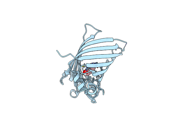
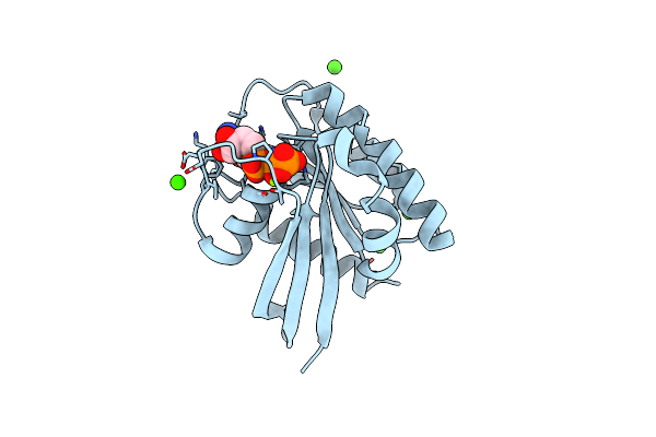
Nitrogen Regulatory Protein Pii From Chlamydomonas Reinhardtii In Unliganded State
Organism: Chlamydomonas reinhardtii
Method: X-RAY DIFFRACTION Resolution:1.60 Å Release Date: 2014-12-03 Classification: SIGNALING PROTEIN Ligands: SO4 |
 |
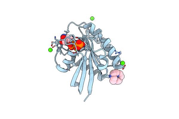
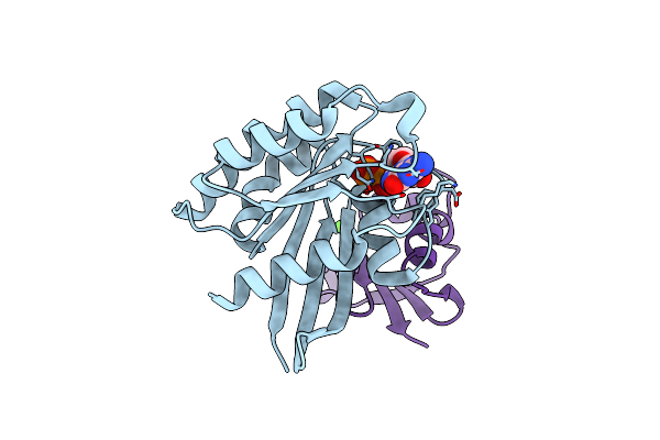
Nitrogen Regulatory Protein Pii From Chlamydomonas Reinhardtii In Complex With Mgatp And 2-Oxoglutarate
Organism: Chlamydomonas reinhardtii
Method: X-RAY DIFFRACTION Resolution:1.45 Å Release Date: 2014-12-03 Classification: SIGNALING PROTEIN Ligands: MG, ATP, AKG, SO4 |
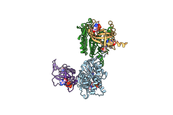 |
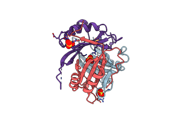
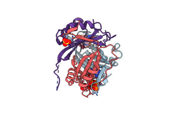
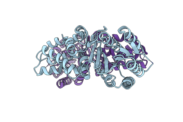
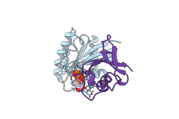
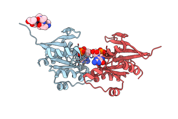
N-Acetylglutamate Kinase From Arabidopsis Thaliana In Complex With Pii From Chlamydomonas Reinhardtii
Organism: Arabidopsis thaliana, Chlamydomonas reinhardtii
Method: X-RAY DIFFRACTION Resolution:2.85 Å Release Date: 2014-12-03 Classification: TRANSFERASE Ligands: ARG, ADP, NLG, MG, X2W, ATP, GLN |
 |
Organism: Guillardia theta ccmp2712
Method: X-RAY DIFFRACTION Resolution:1.95 Å Release Date: 2014-08-13 Classification: LYASE Ligands: HEZ |
 |
Organism: Mus musculus
Method: X-RAY DIFFRACTION Resolution:1.69 Å Release Date: 2013-03-20 Classification: APOPTOSIS Ligands: CD |
 |
Organism: Physcomitrella patens
Method: X-RAY DIFFRACTION Resolution:1.80 Å Release Date: 2013-01-16 Classification: PROTEIN TRANSPORT |
 |
Organism: Homo sapiens
Method: X-RAY DIFFRACTION Resolution:2.02 Å Release Date: 2011-01-05 Classification: ONCOPROTEIN Ligands: GNP, MG, CA, YCN, ZN |
 |
Organism: Homo sapiens
Method: X-RAY DIFFRACTION Resolution:1.44 Å Release Date: 2011-01-05 Classification: ONCOPROTEIN Ligands: GNP, MG, CA |
 |
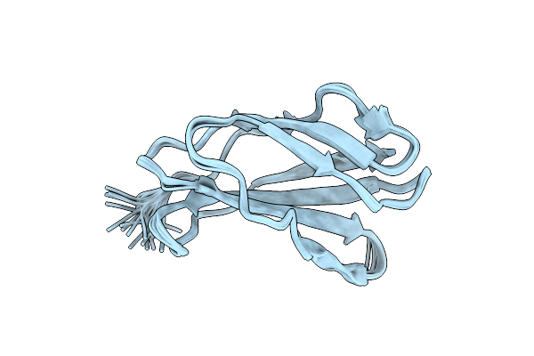
Organism: Rattus norvegicus
Method: SOLUTION NMR Release Date: 2010-08-04 Classification: HYDROLASE Ligands: MG, GDP |
 |
Organism: Homo sapiens
Method: X-RAY DIFFRACTION Resolution:1.92 Å Release Date: 2010-03-23 Classification: GTP binding protein/Transferase Ligands: GDP, MG, CA |
 |
Organism: Homo sapiens
Method: X-RAY DIFFRACTION Resolution:2.15 Å Release Date: 2010-03-23 Classification: GTP BINDING PROTEIN/TRANSFERASE Ligands: GDP, MG |
 |
Organism: Methanocaldococcus jannaschii, Synthetic construct
Method: X-RAY DIFFRACTION Resolution:2.20 Å Release Date: 2009-07-28 Classification: METAL TRANSPORT Ligands: GDP |
 |
Organism: Methanocaldococcus jannaschii
Method: X-RAY DIFFRACTION Resolution:2.10 Å Release Date: 2009-07-28 Classification: METAL TRANSPORT Ligands: GDP, MG, FLC |