
Search Count: 27
 |
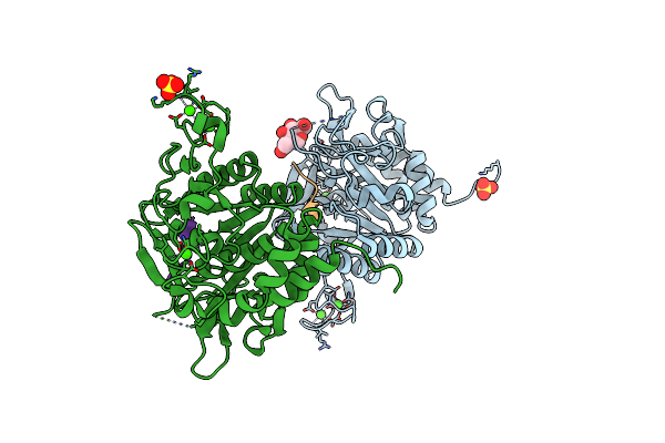
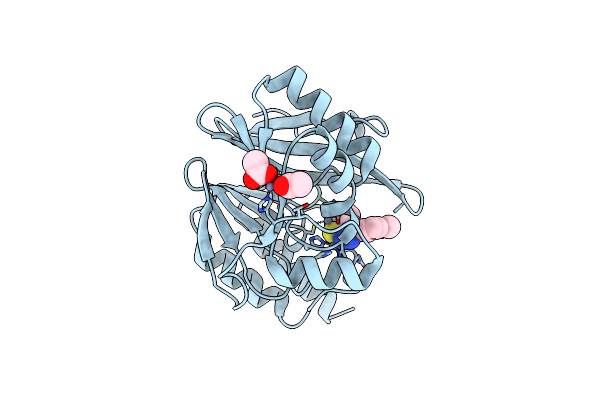
Organism: Plasmodium vivax, Synthetic construct
Method: X-RAY DIFFRACTION Resolution:1.60 Å Release Date: 2025-02-05 Classification: CELL INVASION Ligands: CA, NAG, SO4 |
 |
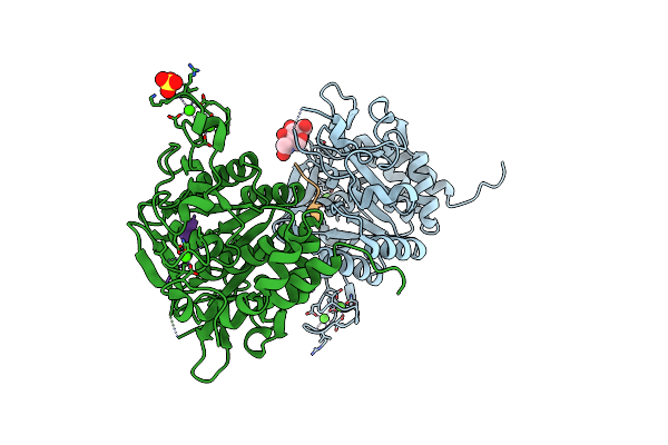
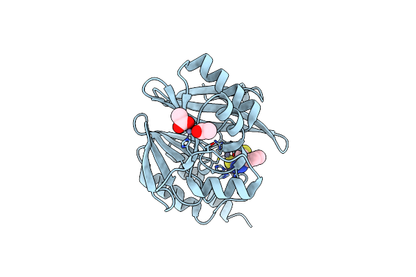
Organism: Plasmodium vivax, Synthetic construct
Method: X-RAY DIFFRACTION Resolution:1.50 Å Release Date: 2024-03-20 Classification: HYDROLASE Ligands: CA, NAG, SO4 |
 |
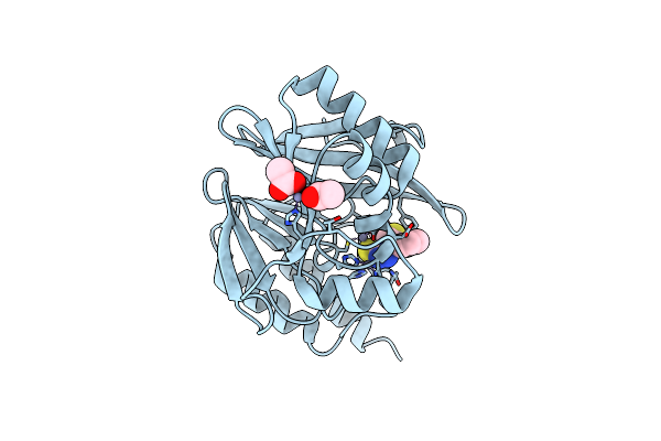
Organism: Plasmodium vivax, Synthetic construct
Method: X-RAY DIFFRACTION Resolution:1.54 Å Release Date: 2024-03-20 Classification: HYDROLASE Ligands: NAG, CA, SO4 |
 |
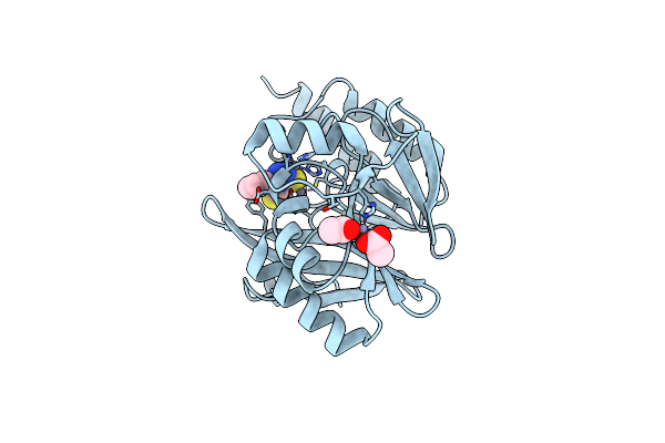
Organism: Plasmodium vivax, Synthetic construct
Method: X-RAY DIFFRACTION Resolution:1.77 Å Release Date: 2024-03-20 Classification: HYDROLASE Ligands: CA, NAG, SO4 |
 |
Structure Of The Catalytic Domain Of P. Vivax Sub1 (Triclinic Crystal Form) In Complex With Inhibitor
Organism: Plasmodium vivax, Synthetic construct
Method: X-RAY DIFFRACTION Resolution:1.51 Å Release Date: 2023-07-19 Classification: HYDROLASE Ligands: NAG, CA, SO4 |
 |
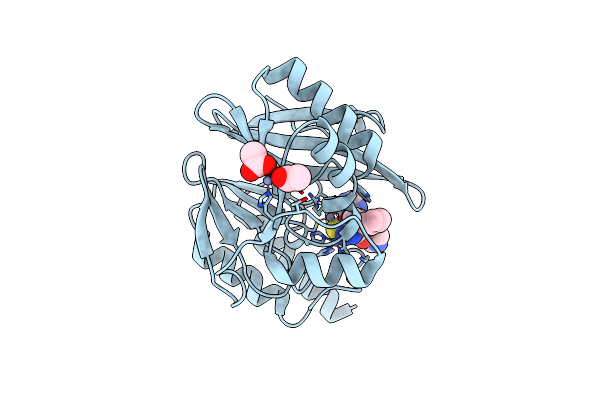
Structure Of The Catalytic Domain Of P. Vivax Sub1 (Triclinic Crystal Form)
Organism: Plasmodium vivax
Method: X-RAY DIFFRACTION Resolution:1.44 Å Release Date: 2023-07-19 Classification: HYDROLASE Ligands: CA, NAG, SO4 |
 |
Organism: Plasmodium vivax
Method: X-RAY DIFFRACTION Resolution:3.25 Å Release Date: 2023-07-19 Classification: HYDROLASE Ligands: CA |
 |
Crystal Structure Of The Vim-2 Acquired Metallo-Beta-Lactamase In Complex With Compound 8 (Jmv-7061)
Organism: Pseudomonas aeruginosa
Method: X-RAY DIFFRACTION Resolution:1.98 Å Release Date: 2023-04-26 Classification: HYDROLASE Ligands: ZN, ACT, L2R |
 |
Metallo-Beta-Lactamase Ndm-1 In Complex With 1,2,4-Triazole-3-Thione Compound 26
Organism: Pseudomonas aeruginosa
Method: X-RAY DIFFRACTION Resolution:1.50 Å Release Date: 2022-12-14 Classification: HYDROLASE Ligands: ZN, CA, L82, EDO, PEG, EPE |
 |
Crystal Structure Of The Vim-2 Acquired Metallo-Beta-Lactamase In Complex With Compound 28 (Jmv-7038)
Organism: Pseudomonas aeruginosa
Method: X-RAY DIFFRACTION Resolution:1.73 Å Release Date: 2022-03-16 Classification: HYDROLASE Ligands: ZN, ACT, 7ZN |
 |
Crystal Structure Of The Vim-2 Acquired Metallo-Beta-Lactamase In Complex With Compound 10 (Jmv-7210)
Organism: Pseudomonas aeruginosa
Method: X-RAY DIFFRACTION Resolution:1.92 Å Release Date: 2021-10-20 Classification: HYDROLASE Ligands: ZN, ACT, UNL |
 |
Crystal Structure Of The Vim-2 Acquired Metallo-Beta-Lactamase In Complex With Compound 8 (Jmv-7207)
Organism: Pseudomonas aeruginosa
Method: X-RAY DIFFRACTION Resolution:1.90 Å Release Date: 2021-10-20 Classification: HYDROLASE Ligands: ZN, ACT, 1TH |
 |
Crystal Structure Of The Vim-2 Acquired Metallo-Beta-Lactamase In Complex With Compound 14 (Jmv-6931)
Organism: Pseudomonas aeruginosa
Method: X-RAY DIFFRACTION Resolution:1.80 Å Release Date: 2021-10-20 Classification: HYDROLASE Ligands: ZN, ACT, UNL |
 |
Crystal Structure Of The Vim-2 Acquired Metallo-Beta-Lactamase In Complex With Jmv-4690 (Cpd 31)
Organism: Pseudomonas aeruginosa
Method: X-RAY DIFFRACTION Resolution:1.95 Å Release Date: 2020-09-30 Classification: HYDROLASE Ligands: ZN, PJB, EDO, ACT, DMS |
 |
1,2,4-Triazole-3-Thione Compounds As Inhibitors Of L1, Di-Zinc Metallo-Beta-Lactamases.
Organism: Stenotrophomonas maltophilia
Method: X-RAY DIFFRACTION Resolution:1.85 Å Release Date: 2017-01-11 Classification: HYDROLASE Ligands: ZN, L3B, SO4 |
 |
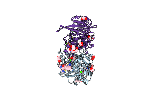
Organism: Plasmodium vivax
Method: X-RAY DIFFRACTION Resolution:2.70 Å Release Date: 2014-09-17 Classification: HYDROLASE Ligands: CA, PO4 |
 |
 |
Solution Structure Of A Linear Analog Of The Cyclic Squash Trypsin Inhibitor Mcoti-Ii
Organism: Momordica cochinchinensis
Method: SOLUTION NMR Release Date: 2007-10-02 Classification: PLANT PROTEIN |
 |
Crystal Structure Of The Zinc-Beta-Lactamase L1 From Stenotrophomonas Maltophilia (Inhibitor 3)
Organism: Stenotrophomonas maltophilia
Method: X-RAY DIFFRACTION Resolution:1.75 Å Release Date: 2007-05-29 Classification: HYDROLASE Ligands: ZN, SO4, L13 |
 |
Crystal Structure Of The Zinc-Beta-Lactamase L1 From Stenotrophomonas Maltophilia (Mono Zinc Form)
Organism: Stenotrophomonas maltophilia
Method: X-RAY DIFFRACTION Resolution:1.80 Å Release Date: 2007-04-17 Classification: HYDROLASE Ligands: ZN, SO4 |