
Search Count: 949
 |
Organism: Saccharomyces cerevisiae (strain atcc 204508 / s288c)
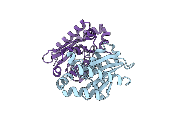
Method: X-RAY DIFFRACTION Release Date: 2025-10-22 Classification: ANTITUMOR PROTEIN Ligands: ZN |
 |
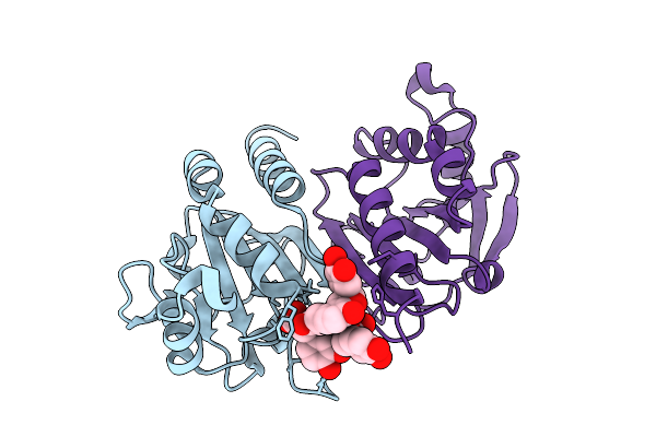
Organism: Human immunodeficiency virus 1
Method: X-RAY DIFFRACTION Release Date: 2025-10-08 Classification: VIRAL PROTEIN Ligands: A1CH4 |
 |
Organism: Human immunodeficiency virus 1
Method: X-RAY DIFFRACTION Release Date: 2025-10-08 Classification: VIRAL PROTEIN Ligands: A1CH5 |
 |
Organism: Human immunodeficiency virus 1
Method: X-RAY DIFFRACTION Release Date: 2025-10-08 Classification: VIRAL PROTEIN Ligands: A1CH6 |
 |
Organism: Human immunodeficiency virus 1
Method: X-RAY DIFFRACTION Release Date: 2025-10-08 Classification: VIRAL PROTEIN Ligands: A1CH7 |
 |
Organism: Homo sapiens
Method: X-RAY DIFFRACTION Release Date: 2025-10-01 Classification: OXIDOREDUCTASE Ligands: A1LXC |
 |
Organism: Synthetic construct
Method: X-RAY DIFFRACTION Release Date: 2025-08-20 Classification: FLUORESCENT PROTEIN Ligands: ABU |
 |
Organism: Methylorubrum extorquens
Method: ELECTRON MICROSCOPY Release Date: 2025-07-23 Classification: PROTEIN BINDING |
 |
Organism: Methylorubrum extorquens
Method: ELECTRON MICROSCOPY Release Date: 2025-07-23 Classification: PROTEIN BINDING Ligands: PQQ |
 |
Organism: Lotus japonicus
Method: X-RAY DIFFRACTION Release Date: 2025-07-16 Classification: PLANT PROTEIN Ligands: NAG, ACT, GOL |
 |
Organism: Medicago truncatula
Method: X-RAY DIFFRACTION Release Date: 2025-07-16 Classification: PLANT PROTEIN Ligands: NAG, SO4, EDO |
 |
Organism: Medicago truncatula
Method: X-RAY DIFFRACTION Release Date: 2025-07-16 Classification: PLANT PROTEIN Ligands: NAG |
 |
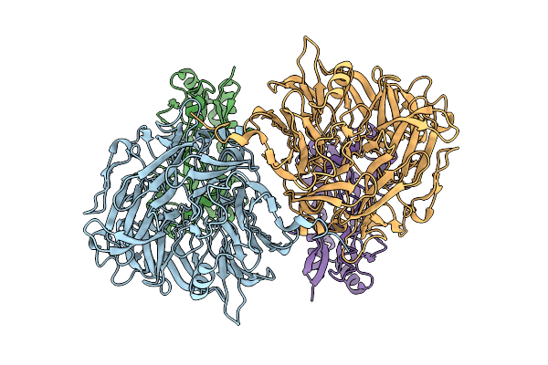
Crystal Structure Of Lotus Japonicus Chip13 Extracellular Domain In Complex With A Nanobody
Organism: Lotus japonicus, Lama glama
Method: X-RAY DIFFRACTION Release Date: 2025-07-16 Classification: PLANT PROTEIN Ligands: NAG |
 |
Crystal Structure Of Lotus Japonicus Chip13 Extracellular Domain In Complex With Chitooctaose
Organism: Lotus japonicus
Method: X-RAY DIFFRACTION Release Date: 2025-07-16 Classification: PLANT PROTEIN Ligands: GOL, NAG |
 |
Organism: Lotus japonicus
Method: X-RAY DIFFRACTION Release Date: 2025-07-16 Classification: PLANT PROTEIN |
 |
Organism: Lotus japonicus, Lama glama
Method: X-RAY DIFFRACTION Release Date: 2025-07-16 Classification: PLANT PROTEIN Ligands: NAG, EDO |
 |
Organism: Lotus japonicus
Method: X-RAY DIFFRACTION Release Date: 2025-07-16 Classification: PLANT PROTEIN Ligands: ACT, IMD, GOL, SO4, NAG |
 |
Crystal Structure Of Lotus Japonicus Chip13 Extracellular Domain In Complex With Chitooctaose
Organism: Lotus japonicus
Method: X-RAY DIFFRACTION Release Date: 2025-07-16 Classification: PLANT PROTEIN Ligands: IMD, EDO |
 |
Organism: Lotus japonicus
Method: X-RAY DIFFRACTION Release Date: 2025-07-16 Classification: PLANT PROTEIN Ligands: NAG, EDO, MAN |
 |
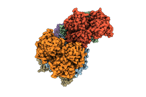
Organism: Homo sapiens
Method: ELECTRON MICROSCOPY Release Date: 2025-06-25 Classification: IMMUNE SYSTEM Ligands: H6P |