Search Count: 31
 |
Crystal Structure Of A Computationally Designed Metalloprotein Containing The Metal-Chelating, Fluorogenic, Non-Canonical Amino Acid 3-(8-Hydroxyquinolin-3-Yl)-L-Alanine: Apo Form
Organism: Saccharolobus solfataricus
Method: X-RAY DIFFRACTION Resolution:1.45 Å Release Date: 2025-02-26 Classification: FLUORESCENT PROTEIN, METAL BINDING PROTEIN Ligands: SO4, GOL |
 |
Crystal Structure Of A Computationally Designed Metalloprotein Containing The Metal-Chelating, Fluorogenic, Non-Canonical Amino Acid 3-(8-Hydroxyquinolin-3-Yl)-L-Alanine: Magnesium-Bound Form
Organism: Saccharolobus solfataricus
Method: X-RAY DIFFRACTION Resolution:1.49 Å Release Date: 2025-02-26 Classification: FLUORESCENT PROTEIN, METAL BINDING PROTEIN Ligands: SO4, MG |
 |
Crystal Structure Of A Computationally Designed Metalloprotein Containing The Metal-Chelating, Fluorogenic, Non-Canonical Amino Acid 3-(8-Hydroxyquinolin-3-Yl)-L-Alanine: Zinc-Bound Form
Organism: Saccharolobus solfataricus
Method: X-RAY DIFFRACTION Resolution:1.45 Å Release Date: 2025-02-26 Classification: FLUORESCENT PROTEIN, METAL BINDING PROTEIN Ligands: SO4, ZN |
 |
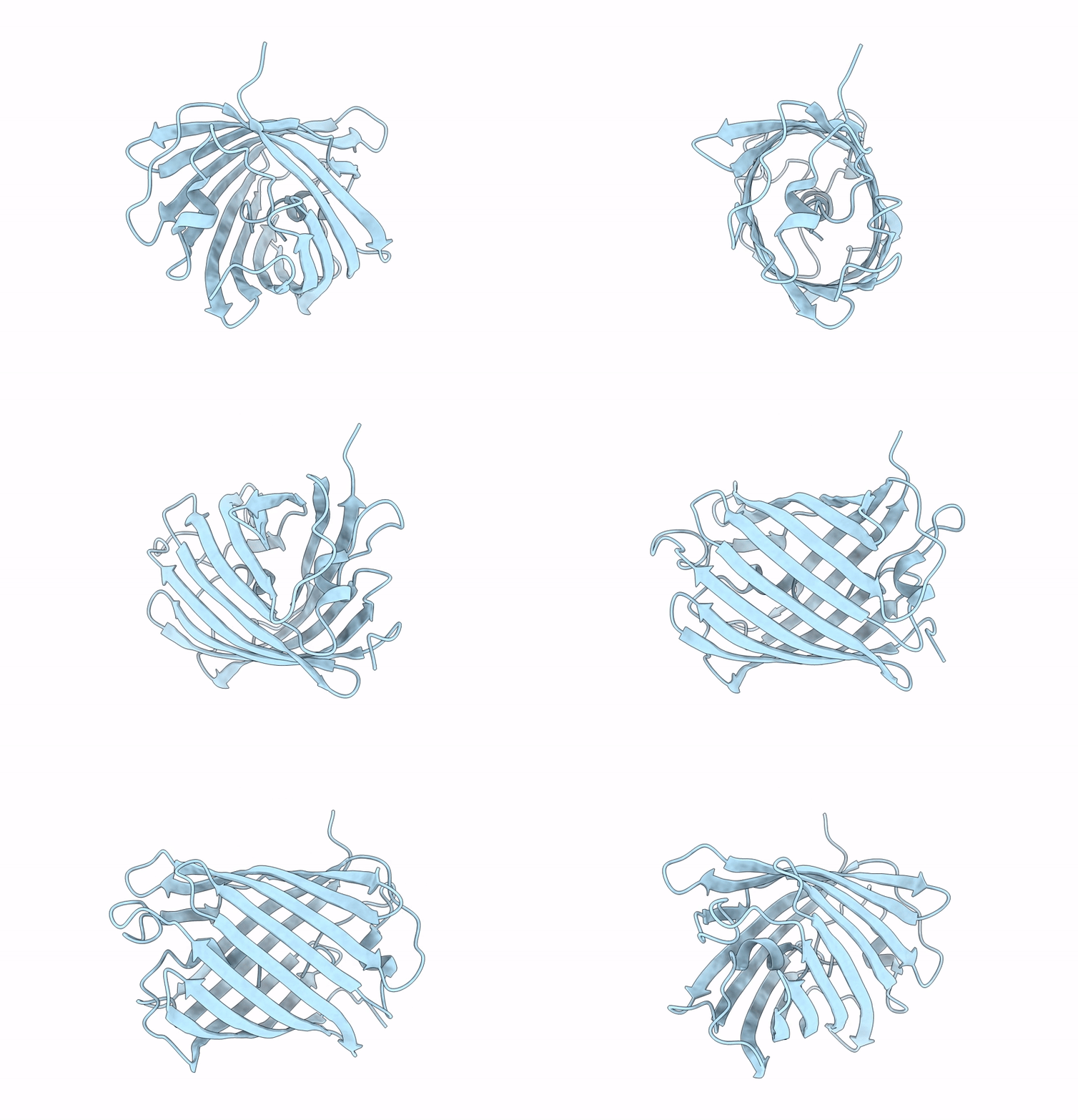
Crystal Structure Of A Reconstructed Kaede-Type Red Fluorescent Protein, Lea A69T
Organism: Synthetic construct
Method: X-RAY DIFFRACTION Resolution:1.50 Å Release Date: 2023-11-29 Classification: FLUORESCENT PROTEIN Ligands: NO3, PEG |
 |
Crystal Structure Of A Reconstructed Kaede-Type Red Fluorescent Protein, Lea H62X, Containing 3-Methylhistidine At Position 62
Organism: Synthetic construct
Method: X-RAY DIFFRACTION Resolution:1.70 Å Release Date: 2023-11-29 Classification: LUMINESCENT PROTEIN |
 |
Crystal Structure Of Fab Containing A Fluorescent Noncanonical Amino Acid With Blocked Excited State Proton Transfer
Organism: Homo sapiens
Method: X-RAY DIFFRACTION Resolution:2.09 Å Release Date: 2022-02-02 Classification: IMMUNE SYSTEM |
 |
Crystal Structure Of A Fab Variant Containing A Fluorescent Noncanonical Amino Acid With Blocked Excited State Proton Transfer And In Complex With Its Antigen, Cd40L
Organism: Homo sapiens
Method: X-RAY DIFFRACTION Resolution:2.00 Å Release Date: 2022-02-02 Classification: CYTOKINE/IMMUNE SYSTEM Ligands: NAG, TRS, PEG, TMO |
 |
Spectroscopic And Structural Characterization Of A Genetically Encoded Direct Sensor For Protein-Ligand Interactions
Organism: Streptomyces avidinii
Method: X-RAY DIFFRACTION Resolution:1.55 Å Release Date: 2021-02-10 Classification: FLUORESCENT PROTEIN Ligands: CL |
 |
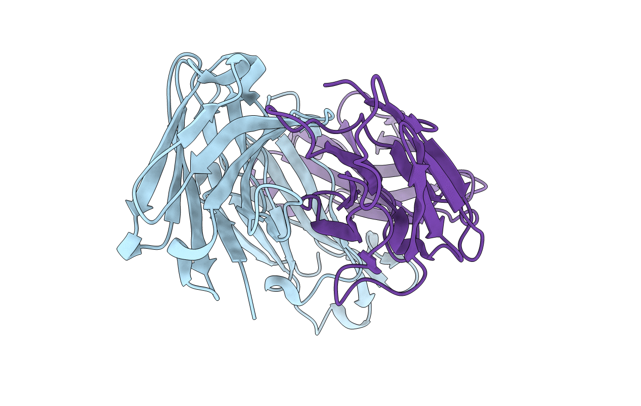
Organism: Homo sapiens
Method: X-RAY DIFFRACTION Resolution:1.77 Å Release Date: 2020-12-23 Classification: IMMUNE SYSTEM |
 |
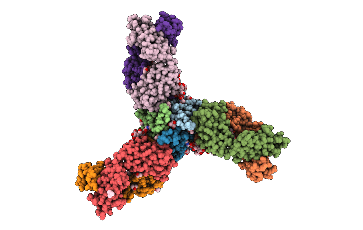
Crystal Structure Of The Fab Fragment Of Humanized 5C8 Antibody Containing The Fluorescent Non-Canonical Amino Acid L-(7-Hydroxycoumarin-4-Yl)Ethylglycine At Ph 9.7
Organism: Homo sapiens
Method: X-RAY DIFFRACTION Resolution:1.75 Å Release Date: 2020-12-23 Classification: IMMUNE SYSTEM |
 |
Crystal Structure Of The Fab Fragment Of Humanized 5C8 Antibody Containing The Fluorescent Non-Canonical Amino Acid L-(7-Hydroxycoumarin-4-Yl)Ethylglycine In Complex With Cd40L At Ph 6.8
Organism: Homo sapiens
Method: X-RAY DIFFRACTION Resolution:1.82 Å Release Date: 2020-12-23 Classification: IMMUNE SYSTEM Ligands: PEG, TRS, TMO, ETE, 1PE |
 |
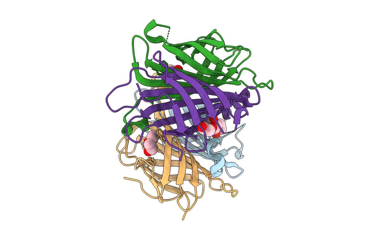
Spectroscopic And Structural Characterization Of A Genetically Encoded Direct Sensor For Protein-Ligand Interactions
Organism: Streptomyces avidinii
Method: X-RAY DIFFRACTION Resolution:1.50 Å Release Date: 2020-09-23 Classification: FLUORESCENT PROTEIN Ligands: GOL, CL |
 |
Spectroscopic And Structural Characterization Of A Genetically Encoded Direct Sensor For Protein-Ligand Interactions
Organism: Streptomyces avidinii
Method: X-RAY DIFFRACTION Resolution:1.55 Å Release Date: 2020-09-23 Classification: FLUORESCENT PROTEIN Ligands: PEG |
 |
Spectroscopic And Structural Characterization Of A Genetically Encoded Direct Sensor For Protein-Ligand Interactions
Organism: Streptomyces avidinii
Method: X-RAY DIFFRACTION Resolution:2.10 Å Release Date: 2020-09-23 Classification: FLUORESCENT PROTEIN Ligands: BTN |
 |
Spectroscopic And Structural Characterization Of A Genetically Encoded Direct Sensor For Protein-Ligand Interactions
Organism: Streptomyces avidinii
Method: X-RAY DIFFRACTION Resolution:1.84 Å Release Date: 2020-09-16 Classification: BIOTIN BINDING PROTEIN Ligands: BTN |
 |
Crystal Structure Of The Fab Fragment Of Humanized 5C8 Antibody Containing The Fluorescent Non-Canonical Amino Acid L-(7-Hydroxycoumarin-4-Yl)Ethylglycine At Ph 5.5
Organism: Homo sapiens
Method: X-RAY DIFFRACTION Resolution:1.45 Å Release Date: 2018-11-14 Classification: IMMUNE SYSTEM |
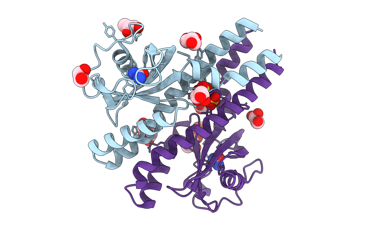 |
Periplasmic Portion Of The Helicobacter Pylori Chemoreceptor Tlpb With Urea Bound
Organism: Helicobacter pylori
Method: X-RAY DIFFRACTION Resolution:1.38 Å Release Date: 2012-06-27 Classification: MEMBRANE PROTEIN Ligands: URE, SO4, PEG, GOL |
 |
Periplasmic Portion Of The Helicobacter Pylori Chemoreceptor Tlpb With Acetamide Bound
Organism: Helicobacter pylori
Method: X-RAY DIFFRACTION Resolution:1.40 Å Release Date: 2012-06-27 Classification: MEMBRANE PROTEIN Ligands: ACM, SO4, GOL |
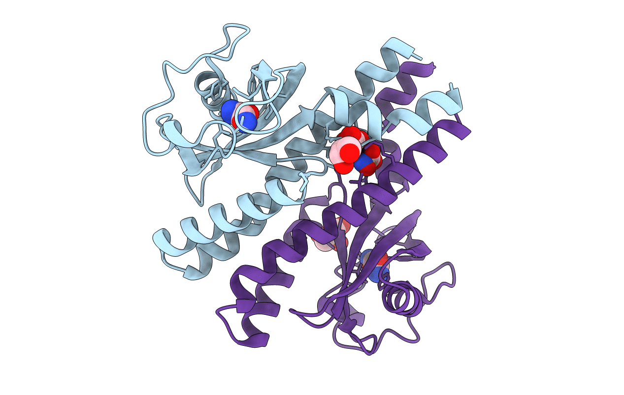 |
Periplasmic Portion Of The Helicobacter Pylori Chemoreceptor Tlpb With Formamide Bound
Organism: Helicobacter pylori
Method: X-RAY DIFFRACTION Resolution:1.42 Å Release Date: 2012-06-27 Classification: MEMBRANE PROTEIN Ligands: ARF, SO4, GOL |
 |
Periplasmic Portion Of The Helicobacter Pylori Chemoreceptor Tlpb With Hydroxyurea Bound
Organism: Helicobacter pylori
Method: X-RAY DIFFRACTION Resolution:1.42 Å Release Date: 2012-06-27 Classification: MEMBRANE PROTEIN Ligands: NHY, SO4, GOL |

