
Search Count: 94
 |
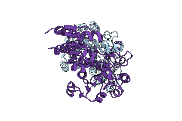
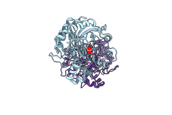
Cryo-Em Structure Of Slc30A10 In Mn2+-Bound State, Determined In Inward-Facing Conformation
Organism: Homo sapiens
Method: ELECTRON MICROSCOPY Release Date: 2025-10-15 Classification: TRANSPORT PROTEIN Ligands: MN |
 |
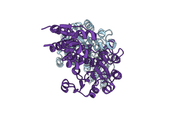
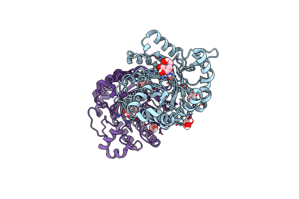
Cryo-Em Structure Of Slc30A10, Determined In Asymmetric Conformations-One Subunit In An Inward-Facing Mn2+-Bound And The Other In An Outward-Facing Mn2+-Unbound Conformation
Organism: Homo sapiens
Method: ELECTRON MICROSCOPY Release Date: 2025-10-15 Classification: TRANSPORT PROTEIN Ligands: MN |
 |
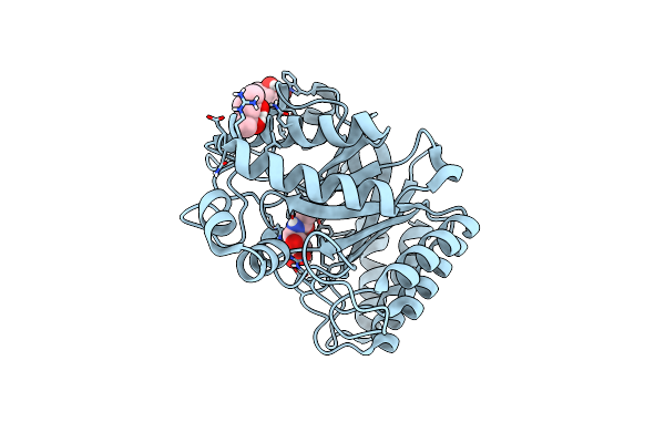
Cryo-Em Structure Of Slc30A10 In The Absence Of Mn2+, Determined In Inward-Facing Conformation
Organism: Homo sapiens
Method: ELECTRON MICROSCOPY Release Date: 2025-10-15 Classification: TRANSPORT PROTEIN |
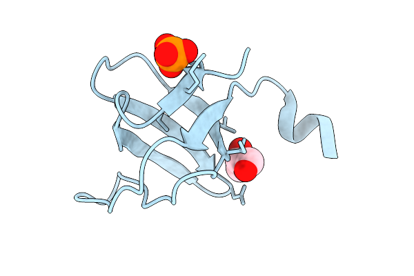 |
Organism: Gallus gallus
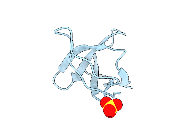
Method: X-RAY DIFFRACTION Release Date: 2025-08-06 Classification: SIGNALING PROTEIN Ligands: GOL, PO4 |
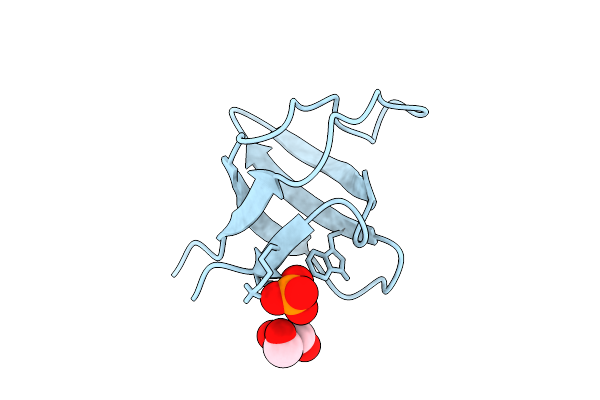 |
Organism: Gallus gallus
Method: X-RAY DIFFRACTION Release Date: 2025-08-06 Classification: SIGNALING PROTEIN Ligands: GOL, PO4 |
 |
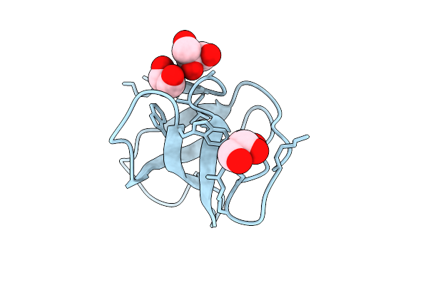
Organism: Gallus gallus
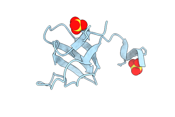
Method: X-RAY DIFFRACTION Release Date: 2025-08-06 Classification: SIGNALING PROTEIN Ligands: GOL, PO4 |
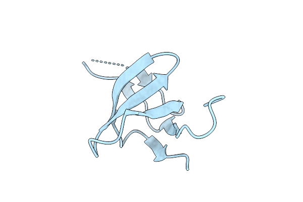 |
Organism: Gallus gallus
Method: X-RAY DIFFRACTION Release Date: 2025-08-06 Classification: SIGNALING PROTEIN |
 |
Organism: Gallus gallus
Method: X-RAY DIFFRACTION Release Date: 2025-08-06 Classification: STRUCTURAL PROTEIN Ligands: GOL |
 |
Organism: Gallus gallus
Method: X-RAY DIFFRACTION Release Date: 2025-08-06 Classification: STRUCTURAL PROTEIN Ligands: SO4 |
 |
Organism: Gallus gallus
Method: X-RAY DIFFRACTION Release Date: 2025-08-06 Classification: STRUCTURAL PROTEIN Ligands: SO4 |
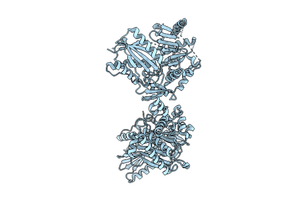 |
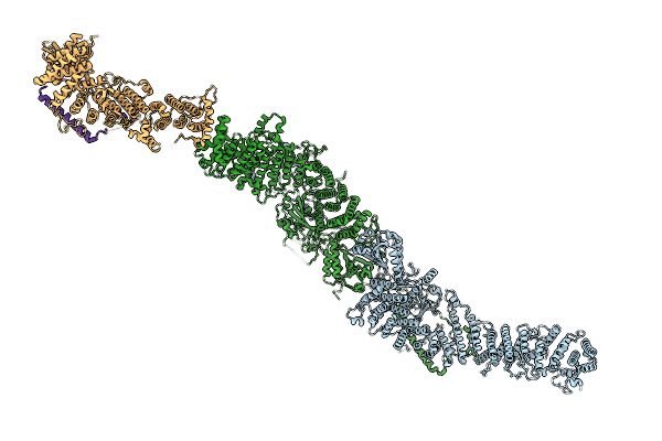
Crystal Structure Of The B. Licheniformis Bacitacin Sythetase 3 Cis-E-Com-C Domains
Organism: Bacillus licheniformis
Method: X-RAY DIFFRACTION Release Date: 2025-08-06 Classification: PEPTIDE BINDING PROTEIN |
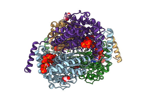 |
Crystal Structure Of Ppk2 Class Iii From Erysipelotrichaceae Bacterium In Complex With Appch2P And Polyphosphate
Organism: Erysipelotrichaceae bacterium
Method: X-RAY DIFFRACTION Release Date: 2025-07-23 Classification: TRANSFERASE Ligands: ACP, 6YY, GOL, BEZ, MG, MPD, A1I4D |
 |
Crystal Structure Of Ppk2 Class Iii From Erysipelotrichaceae Bacterium In Complex With Polyphosphate
Organism: Erysipelotrichaceae bacterium
Method: X-RAY DIFFRACTION Release Date: 2025-07-23 Classification: TRANSFERASE Ligands: 6YY, BEZ, PO4, MG |
 |
Organism: Zea mays
Method: X-RAY DIFFRACTION Release Date: 2025-07-16 Classification: OXIDOREDUCTASE |
 |
Crystal Structure Of Zea Mays 3-Phosphoglycerate Dehydrogenase S282L Mutant
Organism: Zea mays
Method: X-RAY DIFFRACTION Release Date: 2025-07-16 Classification: OXIDOREDUCTASE |
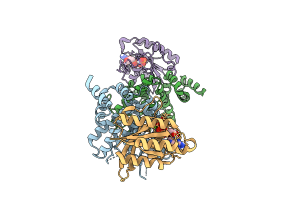 |
Crystal Structure Of The Human Ralgapa2 N-Terminal Domain With Human Kappab-Ras1
Organism: Homo sapiens
Method: X-RAY DIFFRACTION Release Date: 2025-07-16 Classification: SIGNALING PROTEIN Ligands: GNP, MG |
 |
Organism: Homo sapiens
Method: ELECTRON MICROSCOPY Release Date: 2025-07-16 Classification: SIGNALING PROTEIN |
 |
Organism: Terribacillus saccharophilus
Method: X-RAY DIFFRACTION Resolution:1.95 Å Release Date: 2025-03-12 Classification: HYDROLASE Ligands: ACT |
 |
Organism: Terribacillus saccharophilus
Method: X-RAY DIFFRACTION Resolution:1.51 Å Release Date: 2025-03-12 Classification: HYDROLASE Ligands: GC2, MPD |
 |
Organism: Lactiplantibacillus paraplantarum
Method: X-RAY DIFFRACTION Resolution:1.05 Å Release Date: 2025-03-12 Classification: HYDROLASE Ligands: GC2, EDO, PEG |