
Search Count: 4
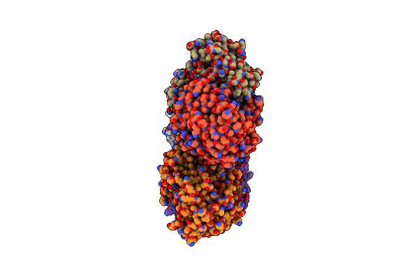 |
Crystal Structure Of Escherichia Coli Enolase Complexed With A Natural Inhibitor Sf2312.
Organism: Escherichia coli
Method: X-RAY DIFFRACTION Resolution:2.24 Å Release Date: 2019-11-27 Classification: LYASE/LYASE inhibitor Ligands: 4NG, MG, GOL, SO4 |
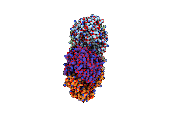 |
Structure Of E.Coli Enolase In Complex With An Analog Of The Natural Product Sf-2312 Metabolite.
Organism: Escherichia coli
Method: X-RAY DIFFRACTION Resolution:2.57 Å Release Date: 2019-11-27 Classification: LYASE Ligands: TLA, SO4, MG, GOL, KVM |
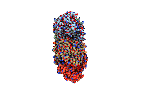 |
Crystal Structure Of Enolase From Escherichia Coli With Bound 2-Phosphoglycerate Substrate
Organism: Escherichia coli
Method: X-RAY DIFFRACTION Resolution:1.81 Å Release Date: 2018-10-31 Classification: LYASE Ligands: 2PG, MG, SO4, GOL |
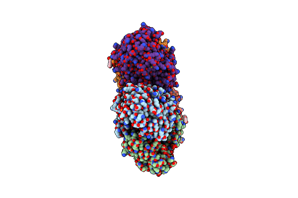 |
Crystal Structure Of Enolase From E. Coli With A Mixture Of Apo Form, Substrate, And Product Form
Organism: Escherichia coli
Method: X-RAY DIFFRACTION Resolution:2.21 Å Release Date: 2018-10-31 Classification: LYASE Ligands: MG, SO4, GOL, PEP, 2PG, MES |