
Search Count: 14
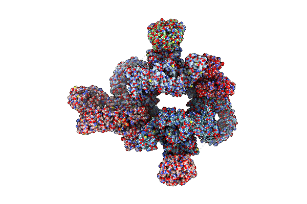 |
Cryoem Structure Of Mtorc1 With A Paediatric Kidney Cancer-Associated 1455-Ewed-1458 Duplication In Mtor, Overall Refinement
Organism: Homo sapiens
Method: ELECTRON MICROSCOPY Release Date: 2024-09-11 Classification: SIGNALING PROTEIN Ligands: ANP, MG |
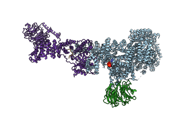 |
Cryoem Structure Of Mtorc1 With A Paediatric Kidney Cancer-Associated 1455-Ewed-1458 Duplication In Mtor, Focused On One Protomer Copy.
Organism: Homo sapiens
Method: ELECTRON MICROSCOPY Release Date: 2024-09-11 Classification: SIGNALING PROTEIN Ligands: ANP, MG |
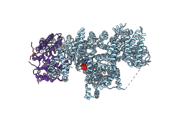 |
Cryoem Structure Of Mtorc1 With A Paediatric Kidney Cancer-Associated 1455-Ewed-1458 Duplication In Mtor, Focused Region Of Mtor And Raptor On One Protomer Copy.
Organism: Homo sapiens
Method: ELECTRON MICROSCOPY Release Date: 2024-09-11 Classification: SIGNALING PROTEIN Ligands: ANP, MG |
 |
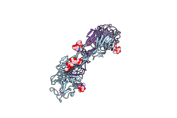
Organism: Homo sapiens
Method: X-RAY DIFFRACTION Resolution:2.89 Å Release Date: 2023-03-15 Classification: CELL ADHESION |
 |
Organism: Homo sapiens
Method: X-RAY DIFFRACTION Resolution:3.09 Å Release Date: 2023-03-15 Classification: CELL ADHESION Ligands: NAG |
 |
Organism: Homo sapiens
Method: X-RAY DIFFRACTION Resolution:3.00 Å Release Date: 2023-03-15 Classification: CELL ADHESION Ligands: NAG, NA |
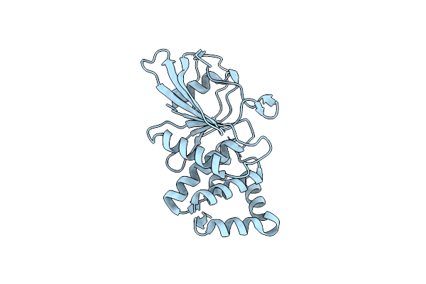 |
Organism: Homo sapiens
Method: X-RAY DIFFRACTION Resolution:1.72 Å Release Date: 2020-07-08 Classification: HYDROLASE Ligands: CL |
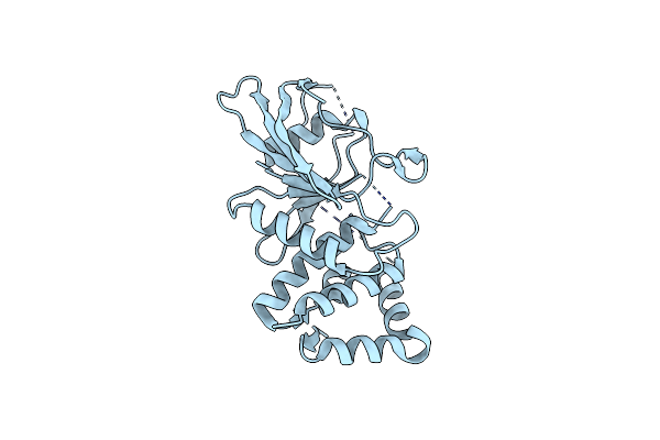 |
Organism: Homo sapiens
Method: X-RAY DIFFRACTION Resolution:1.97 Å Release Date: 2020-07-08 Classification: HYDROLASE |
 |
The Crystal Structure Of The Ferredoxin Protease Fusc In Complex With Its Substrate Plant Ferredoxin
Organism: Pectobacterium atrosepticum scri1043, Arabidopsis thaliana
Method: X-RAY DIFFRACTION Resolution:2.70 Å Release Date: 2018-06-20 Classification: HYDROLASE |
 |
The Crystal Structure Of The Ferredoxin Protease Fusc E83A Mutant In Complex With Arabidopsis Ferredoxin
Organism: Pectobacterium atrosepticum (strain scri 1043 / atcc baa-672), Arabidopsis thaliana
Method: X-RAY DIFFRACTION Resolution:1.90 Å Release Date: 2018-06-20 Classification: HYDROLASE Ligands: ZN |
 |
The Crystal Structure Of The Ferredoxin Protease Fusc In Complex With Arabidopsis Ferredoxin, Ethylmercury Phosphate Soaked Dataset
Organism: Pectobacterium atrosepticum (strain scri 1043 / atcc baa-672), Arabidopsis thaliana
Method: X-RAY DIFFRACTION Resolution:2.30 Å Release Date: 2018-06-20 Classification: HYDROLASE Ligands: PO4, HG |
 |
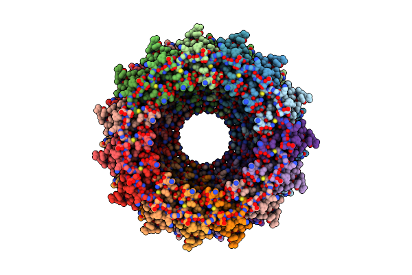
Organism: Escherichia coli o127:h6 (strain e2348/69 / epec)
Method: ELECTRON MICROSCOPY Release Date: 2017-11-15 Classification: MEMBRANE PROTEIN |
 |
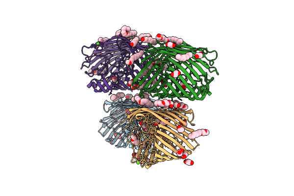
Organism: Pseudomonas aeruginosa (strain atcc 15692 / dsm 22644 / cip 104116 / jcm 14847 / lmg 12228 / 1c / prs 101 / pao1)
Method: ELECTRON MICROSCOPY Release Date: 2017-10-25 Classification: MEMBRANE PROTEIN |
 |
Organism: Pseudomonas aeruginosa
Method: X-RAY DIFFRACTION Resolution:2.30 Å Release Date: 2011-07-27 Classification: TRANSPORT PROTEIN Ligands: CA, EDO, C8E |