
Search Count: 21
 |
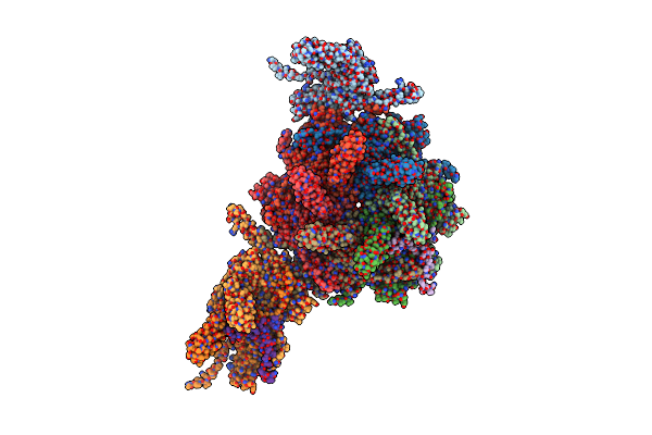
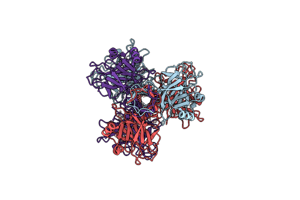
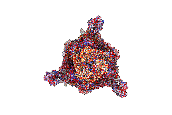
Cryo-Em Structure Of Mycobacteriophage Douge Genome-Packed Vertex (Gp8 And Gp113)
Organism: Mycolicibacterium smegmatis mc2 155
Method: ELECTRON MICROSCOPY Release Date: 2025-07-16 Classification: VIRAL PROTEIN |
 |
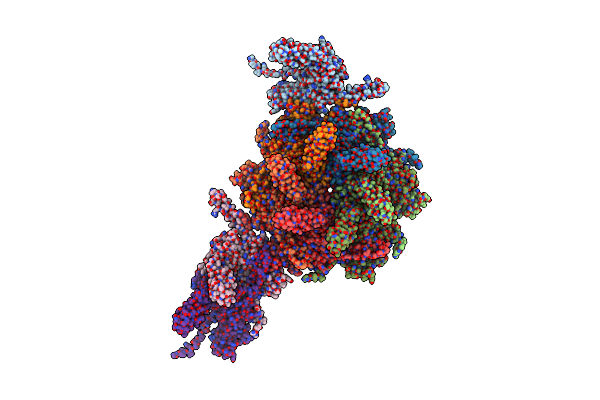
Organism: Mycolicibacterium smegmatis mc2 155
Method: ELECTRON MICROSCOPY Release Date: 2025-07-16 Classification: VIRAL PROTEIN |
 |
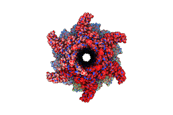
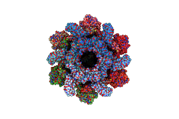
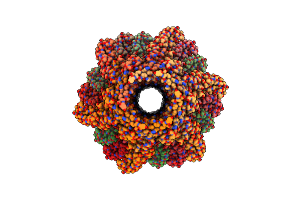
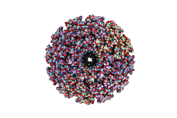
Cryo-Em Structure Of Mycobacteriophage Douge Genome-Packed Capsid (Gp8 And Gp113)
Organism: Mycolicibacterium smegmatis mc2 155
Method: ELECTRON MICROSCOPY Release Date: 2025-06-25 Classification: VIRUS |
 |
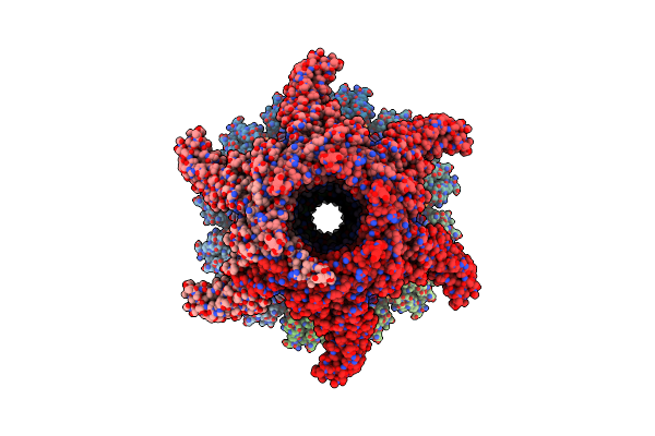
Organism: Mycolicibacterium smegmatis mc2 155
Method: ELECTRON MICROSCOPY Release Date: 2025-06-25 Classification: VIRUS |
 |
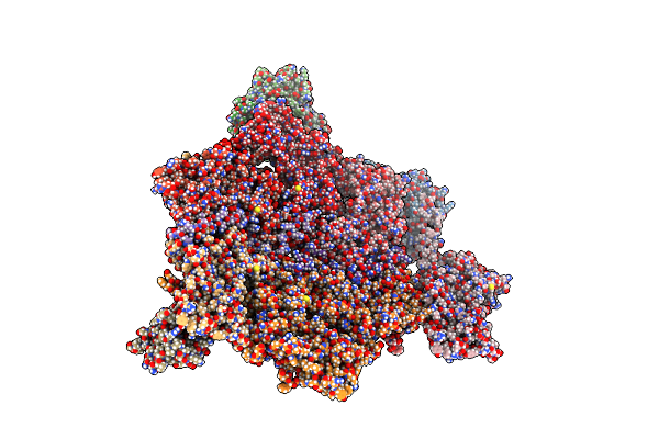
Cryo-Em Structure Of Mycobacteriophage Douge Genome-Packed Connector (Gp5, Gp9, Gp10, Gp12 And Gp13)
Organism: Mycolicibacterium smegmatis mc2 155
Method: ELECTRON MICROSCOPY Release Date: 2025-06-25 Classification: VIRAL PROTEIN |
 |
Cryo-Em Structure Of Mycobacteriophage Douge Genome-Free Connector (Gp5, Gp9, Gp10, Gp12 And Gp13)
Organism: Mycolicibacterium smegmatis mc2 155
Method: ELECTRON MICROSCOPY Release Date: 2025-06-25 Classification: VIRAL PROTEIN |
 |
Organism: Mycolicibacterium smegmatis mc2 155
Method: ELECTRON MICROSCOPY Release Date: 2025-06-25 Classification: VIRAL PROTEIN |
 |
Cryo-Em Structure Of Mycobacteriophage Douge Baseplate (Gp13, Gp17, Gp23, Gp16, Gp18 And Gp20)
Organism: Mycolicibacterium smegmatis mc2 155
Method: ELECTRON MICROSCOPY Release Date: 2025-06-25 Classification: VIRAL PROTEIN |
 |
Organism: Mycolicibacterium smegmatis mc2 155
Method: ELECTRON MICROSCOPY Release Date: 2025-06-25 Classification: VIRAL PROTEIN |
 |
Cryo-Em Structure Of Mycobacteriophage Douge Complete Baseplate (Gp13, Gp17, Gp23, Gp16, Gp18 And Gp20)
Organism: Mycolicibacterium smegmatis mc2 155
Method: ELECTRON MICROSCOPY Release Date: 2025-06-25 Classification: VIRAL PROTEIN |
 |
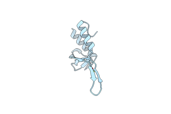
Cryo-Em Structure Of Mycobacteriophage Douge Genome-Packed Tail Tube (Gp13)
Organism: Mycolicibacterium smegmatis mc2 155
Method: ELECTRON MICROSCOPY Release Date: 2025-06-25 Classification: VIRAL PROTEIN |
 |
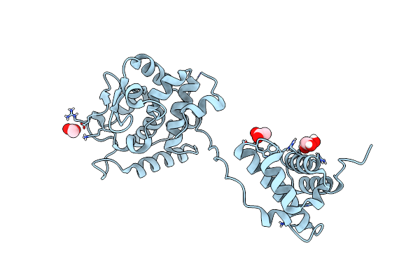
Organism: Mycobacterium phage phaedrus
Method: X-RAY DIFFRACTION Resolution:1.21 Å Release Date: 2024-12-18 Classification: VIRAL PROTEIN Ligands: MG |
 |
Organism: Mycobacterium phage bxb1
Method: ELECTRON MICROSCOPY Release Date: 2024-10-09 Classification: VIRAL PROTEIN |
 |
Organism: Mycobacterium phage bxb1
Method: ELECTRON MICROSCOPY Release Date: 2024-09-25 Classification: VIRAL PROTEIN |
 |
Mycobacteriophage Bxb1 Portal And Connector Assembly - Composite Map And Model
Organism: Mycobacterium phage bxb1
Method: ELECTRON MICROSCOPY Release Date: 2024-09-25 Classification: VIRAL PROTEIN |
 |
Organism: Mycobacterium phage bxb1
Method: ELECTRON MICROSCOPY Release Date: 2024-09-25 Classification: VIRAL PROTEIN |
 |
Organism: Mycobacterium phage bxb1
Method: ELECTRON MICROSCOPY Release Date: 2024-09-25 Classification: VIRAL PROTEIN |
 |
Organism: Mycobacterium phage adjutor
Method: ELECTRON MICROSCOPY Release Date: 2024-04-03 Classification: VIRUS |
 |
Organism: Mycobacterium phage brujita
Method: X-RAY DIFFRACTION Resolution:1.85 Å Release Date: 2018-04-18 Classification: DNA BINDING PROTEIN Ligands: GOL, NA, ACT |
 |
Organism: Mycobacterium phage pukovnik
Method: X-RAY DIFFRACTION Resolution:2.35 Å Release Date: 2013-10-23 Classification: VIRAL PROTEIN Ligands: SO4 |