
Search Count: 39
 |
Organism: Synthetic construct
Method: X-RAY DIFFRACTION Release Date: 2025-08-20 Classification: FLUORESCENT PROTEIN Ligands: ABU |
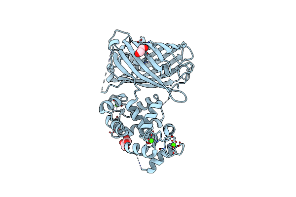 |
Organism: Synthetic construct
Method: X-RAY DIFFRACTION Resolution:2.00 Å Release Date: 2022-11-16 Classification: FLUORESCENT PROTEIN Ligands: CA, GOL, TLA |
 |
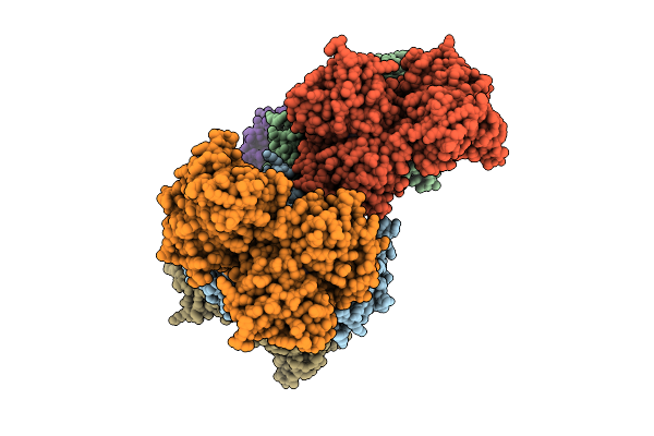
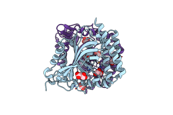
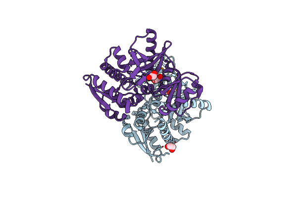
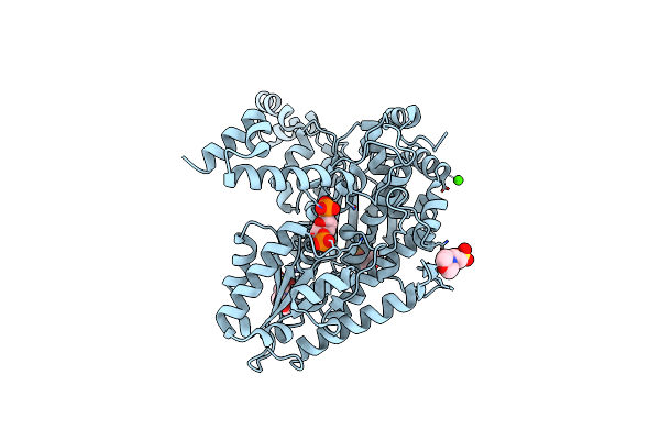
Crystal Structure Of The Flavohem-Like-Fad/Nad Binding Domain Of Nitric Oxide Dioxygenase From Vibrio Cholerae O1 Biovar El Tor
Organism: Vibrio cholerae o1 biovar el tor
Method: X-RAY DIFFRACTION Resolution:2.20 Å Release Date: 2012-04-25 Classification: OXIDOREDUCTASE Ligands: FAD, CL, SO4, GOL |
 |
Organism: Yersinia pestis
Method: X-RAY DIFFRACTION Resolution:1.80 Å Release Date: 2011-04-27 Classification: HYDROLASE Ligands: NO3, FMT |
 |
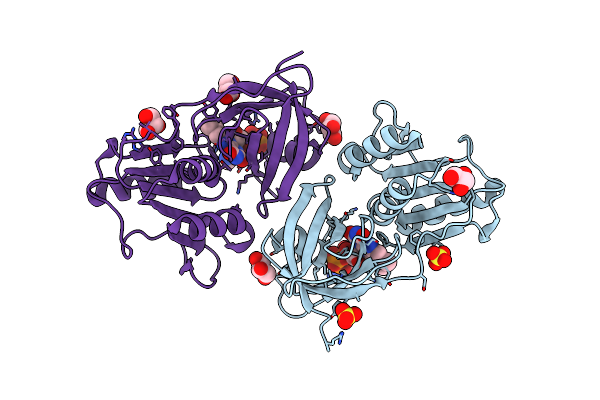
The Crystal Structure Of Glycyl-Trna Synthetase Subunit Alpha From Campylobacter Jejuni Subsp. Jejuni Nctc 11168
Organism: Campylobacter jejuni
Method: X-RAY DIFFRACTION Resolution:2.20 Å Release Date: 2011-04-20 Classification: LIGASE Ligands: LMR, GOL |
 |
Organism: Bacillus anthracis
Method: X-RAY DIFFRACTION Resolution:2.48 Å Release Date: 2011-04-20 Classification: DNA BINDING PROTEIN |
 |
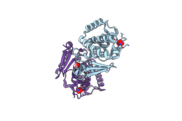
Crystal Structure Of Methionyl-Trna Formyltransferase From Yersinia Pestis Complexed With L-Methionine
Organism: Yersinia pestis
Method: X-RAY DIFFRACTION Resolution:2.26 Å Release Date: 2011-04-13 Classification: TRANSFERASE Ligands: MET, TRS, GOL, MOE |
 |
Crystal Structure Of D-Alanine--D-Alanine Ligase From Bacillus Anthracis Complexed With Atp
Organism: Bacillus anthracis
Method: X-RAY DIFFRACTION Resolution:2.00 Å Release Date: 2011-04-06 Classification: LIGASE Ligands: CA, ATP, MG, EDO, ACY |
 |
Organism: Bacillus anthracis
Method: X-RAY DIFFRACTION Resolution:2.50 Å Release Date: 2011-03-30 Classification: LIGASE Ligands: EDO |
 |
The Crystal Structure Of Ferritin From Vibrio Cholerae O1 Biovar El Tor Str. N16961
Organism: Vibrio cholerae o1 biovar el tor
Method: X-RAY DIFFRACTION Resolution:2.10 Å Release Date: 2011-03-23 Classification: OXIDOREDUCTASE Ligands: EDO |
 |
Crystal Structure Of Phosphoribosylaminoimidazole Synthetase From Francisella Tularensis Complexed With Pyrophosphate
Organism: Francisella tularensis subsp. tularensis
Method: X-RAY DIFFRACTION Resolution:1.80 Å Release Date: 2011-03-16 Classification: LIGASE Ligands: SO4, TRS, POP, PO4, FMT |
 |
Glucose-6-Phosphate Isomerase From Francisella Tularensis Complexed With 6-Phosphogluconic Acid.
Organism: Francisella tularensis subsp. tularensis
Method: X-RAY DIFFRACTION Resolution:1.54 Å Release Date: 2011-02-02 Classification: ISOMERASE Ligands: 6PG, MES, PO4, IPA, CA |
 |
Glucose-6-Phosphate Isomerase From Francisella Tularensis Complexed With Ribose 1,5-Bisphosphate.
Organism: Francisella tularensis subsp. tularensis
Method: X-RAY DIFFRACTION Resolution:1.70 Å Release Date: 2011-01-19 Classification: ISOMERASE Ligands: RI2, MES, PO4, IPA, CA |
 |
Structure Of The Clpp Subunit Of The Atp-Dependent Clp Protease From Coxiella Burnetii
Organism: Coxiella burnetii
Method: X-RAY DIFFRACTION Resolution:2.50 Å Release Date: 2011-01-12 Classification: HYDROLASE Ligands: PEG, CA |
 |
Crystal Structure Of Uridine Phosphorylase Complexed With Uracil From Vibrio Cholerae O1 Biovar El Tor
Organism: Vibrio cholerae o1 biovar el tor
Method: X-RAY DIFFRACTION Resolution:2.00 Å Release Date: 2010-12-15 Classification: TRANSFERASE Ligands: GOL, URA, CL, SO4, FMT, ACY |
 |
Crystal Structure Of Uridine Phosphorylase From Vibrio Cholerae O1 Biovar El Tor
Organism: Vibrio cholerae o1 biovar el tor
Method: X-RAY DIFFRACTION Resolution:1.70 Å Release Date: 2010-10-13 Classification: TRANSFERASE Ligands: GOL, FMT |
 |
Organism: Francisella tularensis subsp. tularensis
Method: X-RAY DIFFRACTION Resolution:1.90 Å Release Date: 2010-10-13 Classification: HYDROLASE Ligands: SO4, GOL |
 |
Organism: Yersinia pestis
Method: X-RAY DIFFRACTION Resolution:1.90 Å Release Date: 2010-08-04 Classification: HYDROLASE Ligands: GOL |
 |
Crystal Structure Of Hexapeptide-Repeat Containing-Acetyltransferase Vca0836 Complexed With Acetyl Co Enzyme A From Vibrio Cholerae O1 Biovar Eltor
Organism: Vibrio cholerae o1 biovar eltor
Method: X-RAY DIFFRACTION Resolution:2.35 Å Release Date: 2010-08-04 Classification: TRANSFERASE Ligands: ACO, ACY, GOL, CL, MG, BU1 |
 |
Crystal Structure Of Phosphoribosylaminoimidazole Synthetase From Francisella Tularensis
Organism: Francisella tularensis subsp. tularensis
Method: X-RAY DIFFRACTION Resolution:1.70 Å Release Date: 2010-07-14 Classification: LIGASE Ligands: SO4, AMP, ACY, FMT, TRS |