Search Count: 131
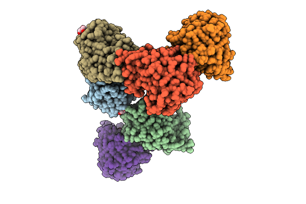 |
Crystal Structure Of Bacteroides Ovatus Kdui1 Responsible For Metabolism Of Glycosaminoglycan
Organism: Bacteroides ovatus atcc 8483
Method: X-RAY DIFFRACTION Release Date: 2025-10-29 Classification: ISOMERASE Ligands: GOL |
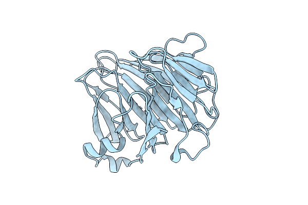 |
Crystal Structure Of Bacteroides Ovatus Kdui2 Responsible For Metabolism Of Glycosaminoglycan
Organism: Bacteroides ovatus atcc 8483
Method: X-RAY DIFFRACTION Release Date: 2025-10-29 Classification: ISOMERASE |
 |
Crystal Structure Of Bacteroides Ovatus Dhud Responsible For Metabolism Of Glycosaminoglycan
Organism: Bacteroides ovatus atcc 8483
Method: X-RAY DIFFRACTION Release Date: 2025-10-29 Classification: OXIDOREDUCTASE Ligands: ACT |
 |
Crystal Structure Of Bacteroides Ovatus Dhud Complexed With Nad+ Responsible For Metabolism Of Glycosaminoglycan
Organism: Bacteroides ovatus atcc 8483
Method: X-RAY DIFFRACTION Release Date: 2025-10-29 Classification: OXIDOREDUCTASE Ligands: NAD |
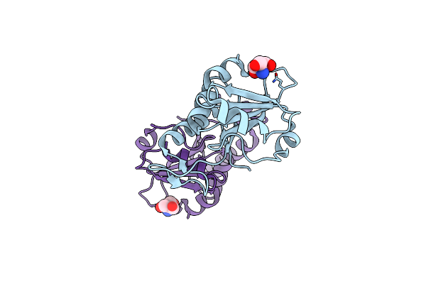 |
Crystal Structure Of Sugar Phosphotransferase System Eiib Component Cpf_0401 From Clostridium Perfringens
Organism: Clostridium perfringens (strain atcc 13124 / dsm 756 / jcm 1290 / ncimb 6125 / nctc 8237 / type a)
Method: X-RAY DIFFRACTION Resolution:1.90 Å Release Date: 2025-04-16 Classification: TRANSFERASE Ligands: TRS |
 |
Crystal Structure Of Lactobacillus Rhamnosus 4-Deoxy-L-Threo-5-Hexosulose-Uronate Ketol-Isomerase Kdui Complexed With Mops
Organism: Lacticaseibacillus rhamnosus
Method: X-RAY DIFFRACTION Resolution:2.80 Å Release Date: 2023-08-16 Classification: ISOMERASE Ligands: ZN, MPO |
 |
Crystal Structure Of Lactobacillus Rhamnosus 4-Deoxy-L-Threo-5-Hexosulose-Uronate Ketol-Isomerase Kdui Complexed With Mes
Organism: Lacticaseibacillus rhamnosus
Method: X-RAY DIFFRACTION Resolution:2.55 Å Release Date: 2023-07-05 Classification: ISOMERASE Ligands: ZN, MES |
 |
Organism: Escherichia coli
Method: X-RAY DIFFRACTION Resolution:1.85 Å Release Date: 2022-12-14 Classification: METAL BINDING PROTEIN Ligands: EDO, ACT, PGE, SO4, ZN |
 |
Crystal Structure Of Lactobacillus Rhamnosus 4-Deoxy-L-Threo-5-Hexosulose-Uronate Ketol-Isomerase Kdui
Organism: Lactobacillus rhamnosus
Method: X-RAY DIFFRACTION Resolution:3.10 Å Release Date: 2022-10-19 Classification: ISOMERASE |
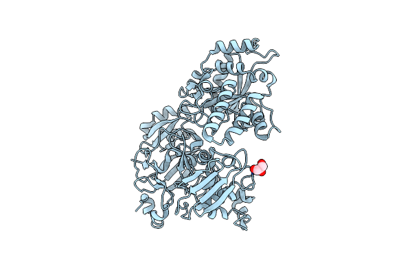 |
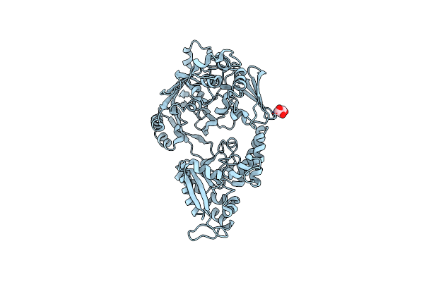
Crystal Structure Of Bacterial Chemotaxis-Dependent Pectin-Binding Protein Sph1118 In An Open Conformation
Organism: Sphingomonas sp. a1
Method: X-RAY DIFFRACTION Resolution:1.70 Å Release Date: 2022-08-17 Classification: SUGAR BINDING PROTEIN Ligands: GOL |
 |
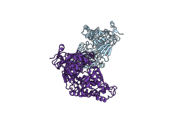
Crystal Structure Of Bacterial Chemotaxis-Dependent Pectin-Binding Protein Sph1118 In A Full Open Conformation
Organism: Sphingomonas sp. a1
Method: X-RAY DIFFRACTION Resolution:1.70 Å Release Date: 2022-08-17 Classification: SUGAR BINDING PROTEIN Ligands: GOL |
 |
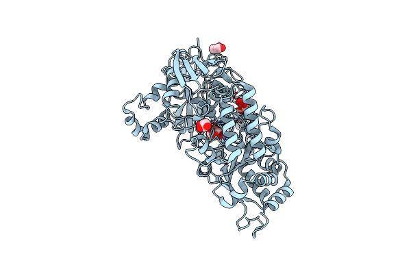
Crystal Structure Of Bacterial Chemotaxis-Dependent Pectin-Binding Protein Sph1118 In A Closed Conformation
Organism: Sphingomonas sp. a1
Method: X-RAY DIFFRACTION Resolution:2.25 Å Release Date: 2022-08-17 Classification: SUGAR BINDING PROTEIN |
 |
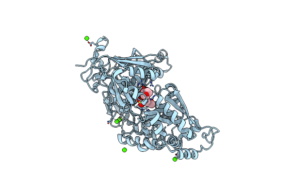
Crystal Structure Of Bacterial Chemotaxis-Dependent Pectin-Binding Protein Sph1118 In Complex With Galacturonic Acid
Organism: Sphingomonas sp. a1
Method: X-RAY DIFFRACTION Resolution:1.74 Å Release Date: 2022-08-17 Classification: SUGAR BINDING PROTEIN Ligands: GOL, ADA |
 |
Crystal Structure Of Bacterial Chemotaxis-Dependent Pectin-Binding Protein Sph1118 In Complex With Mes
Organism: Sphingomonas sp. a1
Method: X-RAY DIFFRACTION Resolution:1.50 Å Release Date: 2022-08-17 Classification: SUGAR BINDING PROTEIN Ligands: CA, MES |
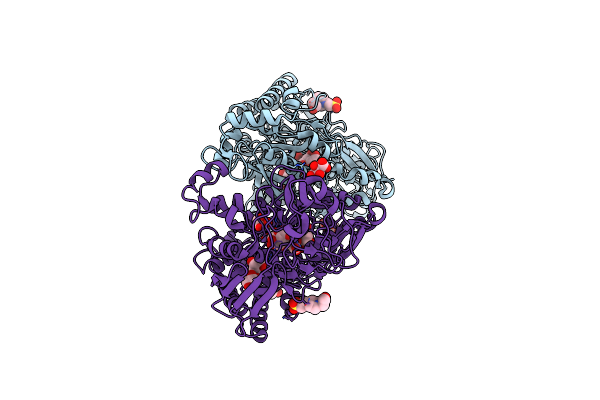 |
Crystal Structure Of Bacterial Chemotaxis-Dependent Pectin-Binding Protein Sph1118 In Complex With Unsaturated Trigalacturonic Acid
Organism: Sphingomonas sp. a1
Method: X-RAY DIFFRACTION Resolution:1.92 Å Release Date: 2022-08-17 Classification: SUGAR BINDING PROTEIN Ligands: EPE, GOL, TLA |
 |
Crystal Structure Of Lactobacillus Rhamnosus 4-Deoxy-L-Threo-5-Hexosulose-Uronate Ketol-Isomerase Kdui Complexed With Hepes
Organism: Lactobacillus rhamnosus
Method: X-RAY DIFFRACTION Resolution:2.79 Å Release Date: 2022-02-23 Classification: ISOMERASE Ligands: EPE, ZN |
 |
Organism: Sphingomonas sp
Method: X-RAY DIFFRACTION Resolution:2.20 Å Release Date: 2020-02-19 Classification: SUGAR BINDING PROTEIN |
 |
Crystal Structure Of Sphingomonas Sp. A1 Peroxidase Efeb Responsible For Import Of Iron
Organism: Sphingomonas sp. a1
Method: X-RAY DIFFRACTION Resolution:2.30 Å Release Date: 2020-01-29 Classification: OXYGEN BINDING Ligands: HEM, OXY, PEG, EDO, PGE |
 |
Organism: Sphingomonas sp. a1
Method: X-RAY DIFFRACTION Resolution:1.88 Å Release Date: 2020-01-29 Classification: TRANSPORT PROTEIN Ligands: EDO, CIT |
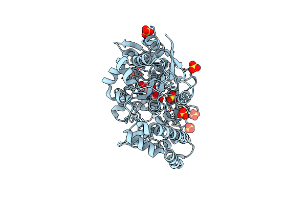 |
Crystal Structure Of Solute-Binding Protein Complexed With Unsaturated Hyaluronan Disaccharide
Organism: Streptobacillus moniliformis dsm 12112
Method: X-RAY DIFFRACTION Resolution:2.29 Å Release Date: 2019-09-11 Classification: SUGAR BINDING PROTEIN Ligands: SO4, CA |

