
Search Count: 19
 |
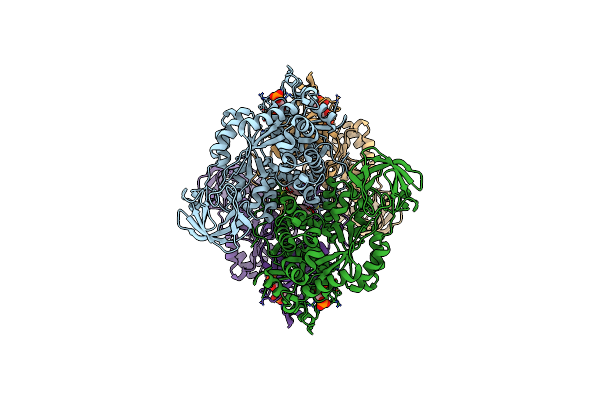
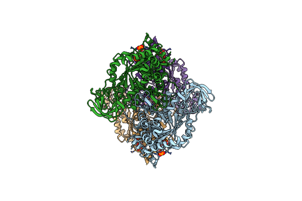
Organism: Escherichia coli
Method: ELECTRON MICROSCOPY Release Date: 2020-01-15 Classification: SUGAR BINDING PROTEIN Ligands: MG, PTQ |
 |
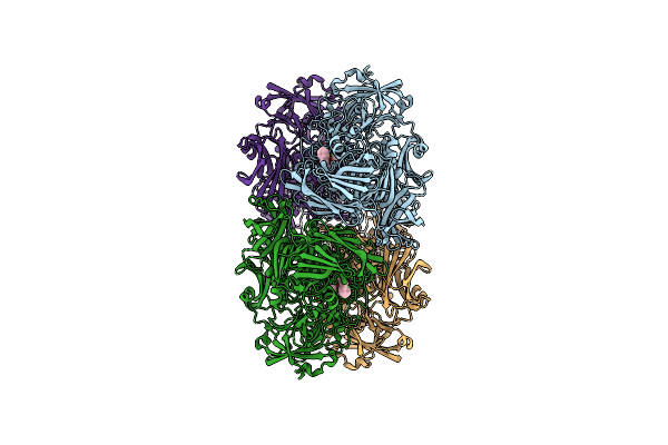
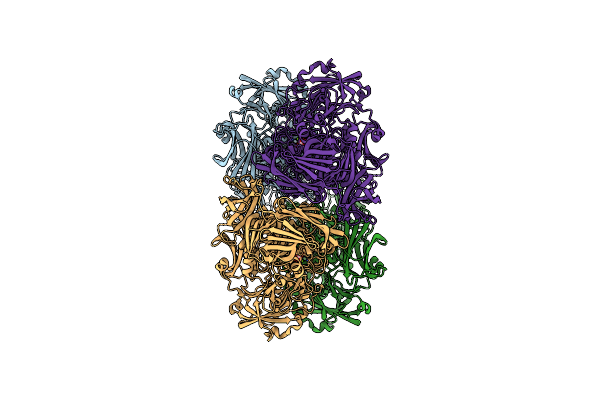
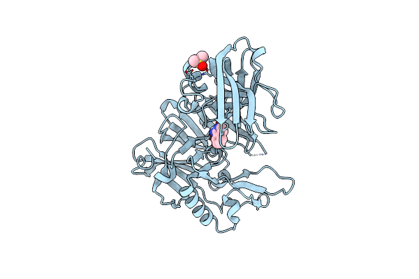
Organism: Homo sapiens
Method: ELECTRON MICROSCOPY Release Date: 2020-01-15 Classification: CYTOSOLIC PROTEIN Ligands: LZ2, FBP |
 |
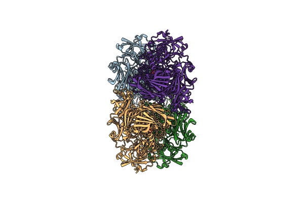
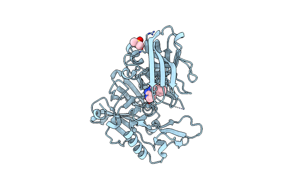
Organism: Homo sapiens
Method: ELECTRON MICROSCOPY Release Date: 2020-01-15 Classification: CYTOSOLIC PROTEIN Ligands: FBP, THR |
 |
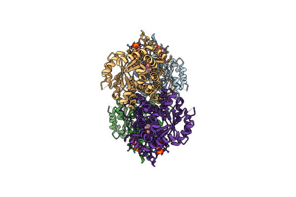
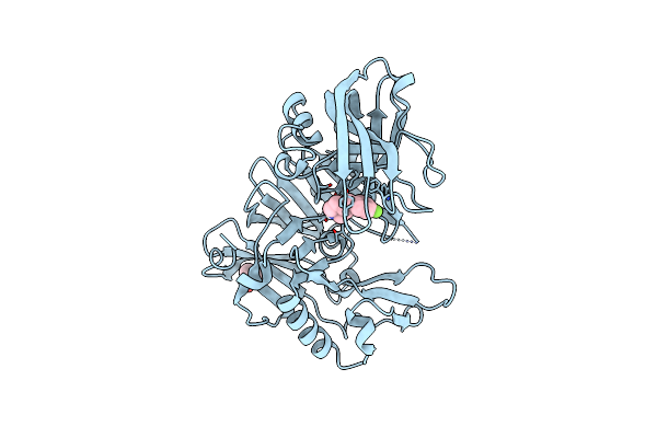
Organism: Homo sapiens
Method: ELECTRON MICROSCOPY Release Date: 2020-01-15 Classification: CYTOSOLIC PROTEIN Ligands: NXE, FBP, DMS |
 |
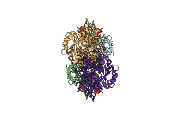
Organism: Homo sapiens
Method: ELECTRON MICROSCOPY Resolution:2.70 Å Release Date: 2020-01-15 Classification: CYTOSOLIC PROTEIN Ligands: NXH, FBP |
 |
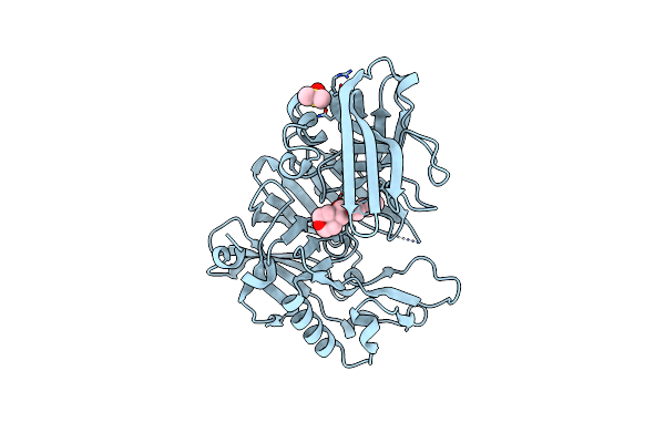
Organism: Escherichia coli
Method: ELECTRON MICROSCOPY Resolution:2.30 Å Release Date: 2020-01-08 Classification: SUGAR BINDING PROTEIN Ligands: MG, DGJ |
 |
Organism: Escherichia coli
Method: ELECTRON MICROSCOPY Resolution:2.30 Å Release Date: 2020-01-08 Classification: SUGAR BINDING PROTEIN Ligands: MG, 0MK |
 |
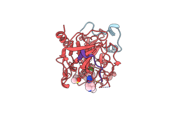
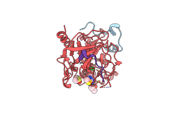
Organism: Homo sapiens
Method: X-RAY DIFFRACTION Resolution:2.25 Å Release Date: 2007-03-13 Classification: HYDROLASE Ligands: IOD, DMS, CMZ |
 |
X-Ray Crystal Structure Of Beta Secretase Complexed With 1-Amino-Isoquinoline
Organism: Homo sapiens
Method: X-RAY DIFFRACTION Resolution:2.20 Å Release Date: 2007-03-13 Classification: HYDROLASE Ligands: IOD, DMS, 1SQ |
 |
Organism: Homo sapiens
Method: X-RAY DIFFRACTION Resolution:2.65 Å Release Date: 2007-03-13 Classification: HYDROLASE Ligands: IOD, DMS, 2AQ |
 |
X-Ray Crystal Structure Of Beta Secretase Complexed With N~3~-Benzylpyridine-2,3-Diamine
Organism: Homo sapiens
Method: X-RAY DIFFRACTION Resolution:2.70 Å Release Date: 2007-03-13 Classification: HYDROLASE Ligands: IOD, DMS, 8AP |
 |
X-Ray Crystal Structure Of Beta Secretase Complexed With 4-(4-Fluorobenzyl)Piperidine
Organism: Homo sapiens
Method: X-RAY DIFFRACTION Resolution:2.15 Å Release Date: 2007-03-13 Classification: HYDROLASE Ligands: IOD, DMS, 4FP |
 |
Organism: Homo sapiens
Method: X-RAY DIFFRACTION Resolution:2.21 Å Release Date: 2005-02-08 Classification: TRANSFERASE Ligands: 3IP |
 |
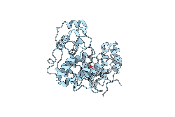
Organism: Homo sapiens
Method: X-RAY DIFFRACTION Resolution:2.20 Å Release Date: 2005-01-27 Classification: HYDROLASE Ligands: MG, LO1 |
 |
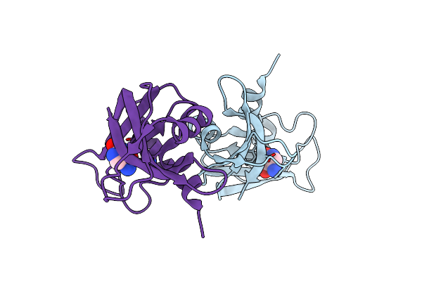
Organism: Hirudo medicinalis, Homo sapiens
Method: X-RAY DIFFRACTION Resolution:2.02 Å Release Date: 2005-01-27 Classification: HYDROLASE/HYDROLASE INHIBITOR Ligands: L02, DMS |
 |
Organism: Hirudo medicinalis, Homo sapiens
Method: X-RAY DIFFRACTION Resolution:2.20 Å Release Date: 2005-01-27 Classification: HYDROLASE/HYDROLASE INHIBITOR Ligands: L03, DMS |
 |
Organism: Homo sapiens
Method: X-RAY DIFFRACTION Resolution:2.16 Å Release Date: 2005-01-27 Classification: TRANSFERASE Ligands: 2CH |
 |
Organism: Bos taurus
Method: X-RAY DIFFRACTION Resolution:1.90 Å Release Date: 2005-01-27 Classification: HYDROLASE Ligands: WBU |
 |
Organism: Homo sapiens
Method: X-RAY DIFFRACTION Resolution:2.20 Å Release Date: 2005-01-27 Classification: TRANSFERASE Ligands: CIG |