
Search Count: 81
 |
Organism: Mycolicibacterium smegmatis
Method: X-RAY DIFFRACTION Resolution:2.00 Å Release Date: 2023-07-05 Classification: CYTOSOLIC PROTEIN Ligands: RPI |
 |
Organism: Mycobacterium tuberculosis (strain atcc 25618 / h37rv), Nonomuraea sp. mjm5123
Method: X-RAY DIFFRACTION Resolution:2.25 Å Release Date: 2023-07-05 Classification: CHAPERONE Ligands: FMT |
 |
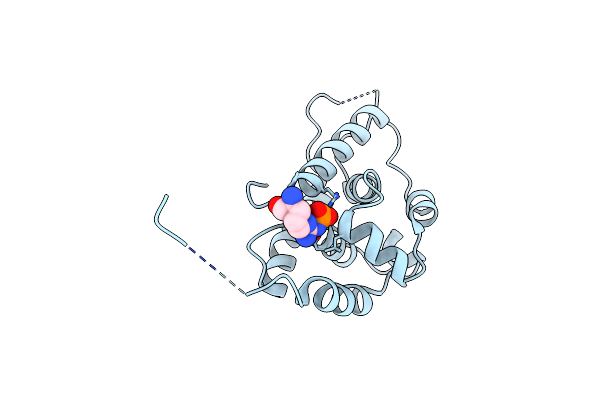
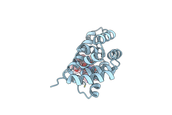
Crystal Structure Of The Carbonic Anhydrase-Like Domain Of Ccmm From Synechococcus Elongatus (Strain Pcc 7942)
Organism: Synechococcus elongatus (strain pcc 7942 / fachb-805)
Method: X-RAY DIFFRACTION Resolution:1.67 Å Release Date: 2021-11-10 Classification: PHOTOSYNTHESIS Ligands: NI, CL |
 |
Crystal Structure Of The Carbonic Anhydrase-Like Domain Of Ccmm In Complex With The C-Terminal 17 Residues Of Ccaa From Synechococcus Elongatus (Strain Pcc 7942)
Organism: Synechococcus elongatus (strain pcc 7942 / fachb-805)
Method: X-RAY DIFFRACTION Resolution:1.63 Å Release Date: 2021-11-10 Classification: PHOTOSYNTHESIS Ligands: NI, CL |
 |
Cryo-Em Structure Of B. Subtilis Clpc (Dwb Mutant) Hexamer Bound To A Substrate Polypeptide
Organism: Bacillus subtilis (strain 168), Unidentified
Method: ELECTRON MICROSCOPY Release Date: 2021-10-06 Classification: CHAPERONE Ligands: ADP, ATP |
 |
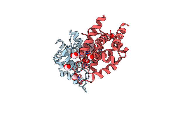
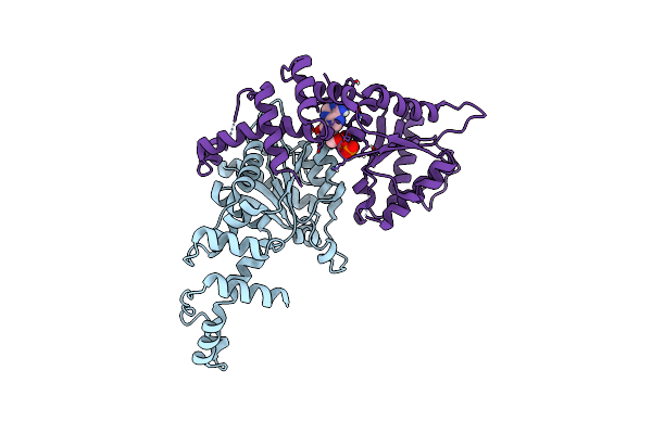
Structure Of Autoinhibited Akt1 Reveals Mechanism Of Pip3-Mediated Activation
Organism: Homo sapiens, Danio rerio, Lama glama
Method: X-RAY DIFFRACTION Resolution:2.05 Å Release Date: 2021-08-25 Classification: SIGNALING PROTEIN |
 |
Organism: Mycobacterium tuberculosis, Streptomyces
Method: X-RAY DIFFRACTION Resolution:1.68 Å Release Date: 2021-08-11 Classification: CHAPERONE |
 |
Organism: Homo sapiens
Method: X-RAY DIFFRACTION Resolution:3.20 Å Release Date: 2021-05-12 Classification: OXIDOREDUCTASE Ligands: FAD |
 |
Crystal Structure Of The Aaa Domain Of Rubisco Activase From Nostoc Sp. (Strain Pcc 7120), Gadolinium Complex
Organism: Nostoc sp. pcc 7120 = fachb-418
Method: X-RAY DIFFRACTION Resolution:2.71 Å Release Date: 2020-09-23 Classification: CHAPERONE Ligands: GD, CL, ADP |
 |
Crystal Structure Of The Aaa Domain Of Rubisco Activase From Nostoc Sp. (Strain Pcc 7120)
Organism: Nostoc sp. pcc 7120 = fachb-418
Method: X-RAY DIFFRACTION Resolution:2.45 Å Release Date: 2020-09-23 Classification: CHAPERONE Ligands: CL, ADP |
 |
Cryoem Structure Of Rubisco Activase With Its Substrate Rubisco From Nostoc Sp. (Strain Pcc7120)
Organism: Nostoc sp. (strain pcc 7120 / sag 25.82 / utex 2576)
Method: ELECTRON MICROSCOPY Release Date: 2020-09-23 Classification: CHAPERONE Ligands: ADP, AGS, MG, CAP |
 |
Cryoem Structure Of The Interaction Between Rubisco Activase Small-Subunit-Like (Ssul) Domain With Rubisco From Nostoc Sp. (Strain Pcc7120)
Organism: Nostoc sp. (strain pcc 7120 / sag 25.82 / utex 2576)
Method: ELECTRON MICROSCOPY Release Date: 2020-09-23 Classification: PHOTOSYNTHESIS |
 |
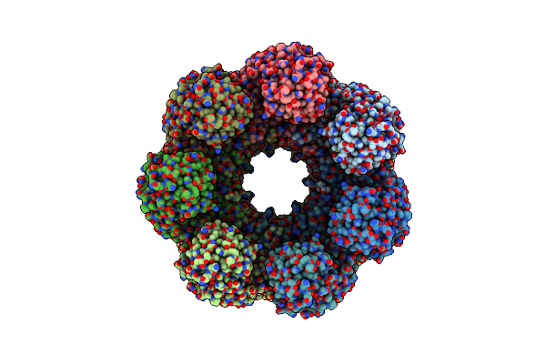
Crystal Structure Of The Chaperonin Gp146 From The Bacteriophage El 2 (Pseudomonas Aeruginosa) In Presence Of Atp-Befx, Crystal Form I
Organism: Pseudomonas phage el
Method: X-RAY DIFFRACTION Resolution:4.03 Å Release Date: 2020-04-22 Classification: CHAPERONE Ligands: MG, ATP |
 |
Crystal Structure Of The Chaperonin Gp146 From The Bacteriophage El 2 (Pseudomonas Aeruginosa) In Presence Of Atp-Befx, Crystal Form Ii
Organism: Pseudomonas phage el
Method: X-RAY DIFFRACTION Resolution:3.54 Å Release Date: 2020-04-22 Classification: CHAPERONE Ligands: MG, K, ATP, ADP |
 |
Structure Of The Chaperonin Gp146 From The Bacteriophage El (Pseudomonas Aeruginosa) In The Apo State
Organism: Pseudomonas phage el
Method: ELECTRON MICROSCOPY Release Date: 2020-04-22 Classification: CHAPERONE |
 |
Structure Of The Chaperonin Gp146 From The Bacteriophage El (Pseudomonas Aeruginosa) In Complex With Adp
Organism: Pseudomonas phage el
Method: ELECTRON MICROSCOPY Release Date: 2020-04-22 Classification: CHAPERONE Ligands: ADP |
 |
Structure Of The Chaperonin Gp146 From The Bacteriophage El (Pseudomonas Aeruginosa) In Complex With Atpgammas
Organism: Pseudomonas phage el
Method: ELECTRON MICROSCOPY Release Date: 2020-04-22 Classification: CHAPERONE Ligands: MG, K, AGS |
 |
Crystal Structure Of The Small Subunit-Like Domain Of Rubisco Activase From Nostoc Sp. (Strain Pcc 7120)
Organism: Nostoc sp. (strain pcc 7120 / sag 25.82 / utex 2576)
Method: X-RAY DIFFRACTION Resolution:1.38 Å Release Date: 2020-01-15 Classification: CHAPERONE Ligands: NI |
 |
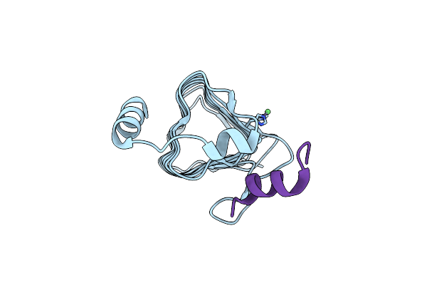
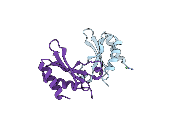
A Ubiquitin-Like Dimerization Domain Controls Protein Kinase D Activation By Trans-Autophosphorylation
Organism: Caenorhabditis elegans
Method: X-RAY DIFFRACTION Resolution:2.26 Å Release Date: 2019-08-14 Classification: SIGNALING PROTEIN Ligands: ZN |
 |
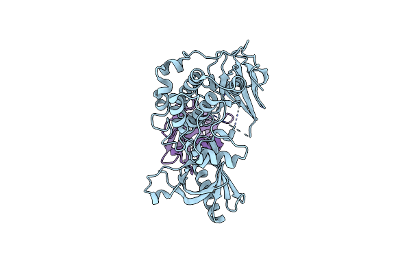
Crystal Structure Of Redox-Inhibited Phosphoribulokinase From Synechococcus Sp. (Strain Pcc 6301)
Organism: Synechococcus sp. (strain atcc 27144 / pcc 6301 / saug 1402/1)
Method: X-RAY DIFFRACTION Resolution:2.40 Å Release Date: 2019-03-27 Classification: TRANSFERASE |