Planned Maintenance: Some services may turn out to be unavailable from 15th January, 2026 to 16th January, 2026. We apologize for the inconvenience!

Planned Maintenance: Some services may turn out to be unavailable from 15th January, 2026 to 16th January, 2026. We apologize for the inconvenience!
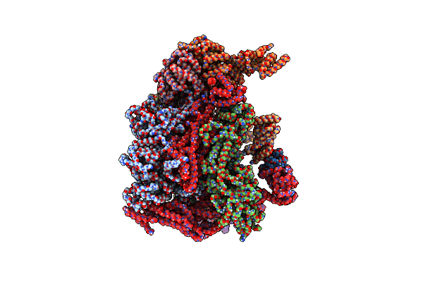 |
Organism: Homo sapiens
Method: ELECTRON MICROSCOPY Release Date: 2023-09-27 Classification: TRANSFERASE Ligands: ZN |
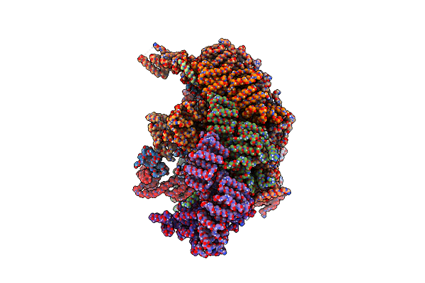 |
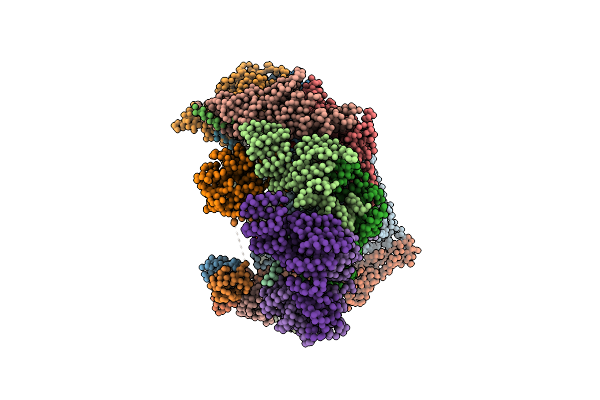
Organism: Homo sapiens
Method: ELECTRON MICROSCOPY Release Date: 2023-09-27 Classification: LIGASE Ligands: ZN |
 |
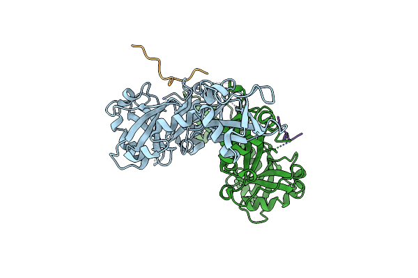
Organism: Homo sapiens, Human betaherpesvirus 5
Method: X-RAY DIFFRACTION Resolution:2.90 Å Release Date: 2022-08-10 Classification: PROTEIN BINDING |
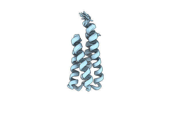 |
Organism: Synthetic construct
Method: SOLUTION NMR Release Date: 2022-03-02 Classification: DE NOVO PROTEIN |
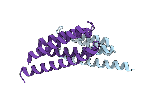 |
Organism: Synthetic construct
Method: X-RAY DIFFRACTION Resolution:1.34 Å Release Date: 2022-03-02 Classification: DE NOVO PROTEIN |
 |
Model Of Human Anaphase-Promoting Complex/Cyclosome (Apc/C-Cdh1) With E2 Ube2S Poised For Polyubiquitination Where Ube2S, Apc2, And Apc11 Are Modeled Into Low Resolution Density
Organism: Homo sapiens, Saccharomyces cerevisiae (strain atcc 204508 / s288c)
Method: ELECTRON MICROSCOPY Release Date: 2016-10-26 Classification: CELL CYCLE |
 |
Model Of Human Anaphase-Promoting Complex/Cyclosome (Apc/C-Cdh1) With A Cross Linked Ubiquitin Variant-Substrate-Ube2C (Ubch10) Complex Representing Key Features Of Multiubiquitination
Organism: Homo sapiens, Saccharomyces cerevisiae, Rattus norvegicus
Method: ELECTRON MICROSCOPY Release Date: 2016-09-14 Classification: SIGNALING PROTEIN |
 |
Apc11-Ubv Shows Role Of Noncovalent Ring-Ubiquitin Interactions In Processive Multiubiquitination And Ubiquitin Chain Elongation By Apc/C
Organism: Homo sapiens
Method: X-RAY DIFFRACTION Resolution:2.00 Å Release Date: 2016-06-15 Classification: CELL CYCLE Ligands: ZN |
 |
Organism: Homo sapiens
Method: X-RAY DIFFRACTION Resolution:2.29 Å Release Date: 2015-09-09 Classification: HYDROLASE/LIGASE |
 |
Structure Of An Rsp5Xubxsna3 Complex: Mechanism Of Ubiquitin Ligation And Lysine Prioritization By A Hect E3
Organism: Saccharomyces cerevisiae, Homo sapiens
Method: X-RAY DIFFRACTION Resolution:3.10 Å Release Date: 2013-08-14 Classification: Ligase/protein binding |
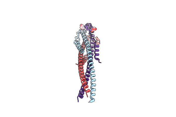 |
Crystal Structure Of The Marburg Virus Gp2 Ectodomain In Its Post-Fusion Conformation
Organism: Saccharomyces cerevisiae, Lake victoria marburgvirus
Method: X-RAY DIFFRACTION Resolution:1.90 Å Release Date: 2012-09-12 Classification: VIRAL PROTEIN Ligands: GOL, CL |